Download
1 / 36
400 likes | 945 Vues
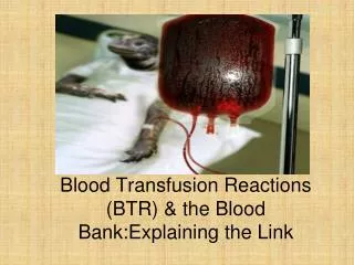
Blood Transfusion Reactions (BTR) & the Blood Bank:Explaining the Link. Objectives. Early identification of common transfusion reactions Differentiate life threatening reactions from benign transfusion reactions Manage common immunologic transfusion reactions. Febrile non hemolytic tranx rxns

E N D
Blood Transfusion Reactions(BTR) & the Blood Bank:Explaining the Link
Objectives • Early identification of common transfusion reactions • Differentiate life threatening reactions from benign transfusion reactions • Manage common immunologic transfusion reactions.
Febrile non hemolytic tranx rxns Immune mediated hemolysis ---Acute and delayed hemolytic reactions Anaphylactic transfusion rxns Urticarial transfusion rxns Post-transfusion purpura GVHD TRALI –Transfusion Related Acute Lung Injury (pulm leuko-agglutinin reactions) Immune mediated transfusion reactions
Non immune mediated reactions • Physical reactions: thermal i.e. heat or cold induced • Infectious; Hepatitis B/C, malaria, HIV, etc. • Chemical; citrate toxicity, hypo/hyperkalemia, iron overload • Acute hypotensive reaction: mediated by bradykinins and occurs in patients with faulty bradykinin metabolism on ACE I • Osmotic injury • Congenital and acquired hemolytic anemias
What to DO ? • Early recognition of signs/symptoms suggestive of a transfusion reaction and prompt reporting to the blood bank are essential. • The most common symptoms are chills, rigors, fever, dyspnea, light-headedness, urticaria, itching, and flank pain. If any of these symptoms (other than localized urticaria and itching) (?) occur, the transfusion should be stopped immediately and the IV line kept open with normal saline.
What to DO? • The remainder of the blood product and clotted and anticoagulated samples of the patient's blood should be sent to the blood bank for investigation.
Immunologic Reactionsclassic blood transfusion reaction are usually immunologic and occur to interactions of inherited/ acquired Ab with foreign Ag from transfused blood Incidence of rxns -most common cause is transfused of non-matched blood mostly to clerical error -2x more common in infants than adults -more common in patients with hematological and oncological conditions
Febrile Non Hemolytic Tranfusion Reactions(FNHTR) • Most common, usually benign without sequela • A least rise of temp. of 1o C • 15% will have a reaction in the future with subsequent transfusion Etiology • Class 1 HLA Ab (HLA 5)directed against contaminating WBC in red cell concentration • Cytokines IL-1, 6,8 and TNF alpha generated in stored blood/products
Management Discontinue transfusion, rule out hemolysis i.e. check labels, repeat type and cross, coombs test Prevention • Leukoreduction
Acute Hemolytic Reactions • Rapid destruction of RBC immediately after within 24 hours of transfusion. • Occurs due to rapid transfused RBC destruction by preformed recipients Abs • IgM mediated complement fixation leading to rapid intra vascular hemolysis • Most common causes are clerical or procedural errors
Acute hemolytic rxns • Clinical presentation • Classic presenting triad of Fever, flank pain and reddish brown urine from hemoglobinuria. • DIC may be presenting mode
Conditions – destroy donor cells • Naturally occuring or stimulated alloantibodies (anti-A, Kell, Jka , Fya) • Autoantibodies • Drug-associated antibodies • Bacterial Contamination
Conditions –destroy recipient cells • Incompatible (ABO) • Infusion of large amount of hypotonic solutions • Mechanical Trauma
Actions ? • If AHTR is suspected, one of the first steps is to recheck the sample and patient identifications. Diagnosis is confirmed by measuring urinary Hb, bilirubin, and haptoglobin. • Intravascular hemolysis produces free Hb in the plasma and urine; haptoglobin levels are very low. Hyperbilirubinemia may follow. • After the acute phase, the degree of acute renal failure determines the prognosis. • Prolonged oliguria and shock are poor prognostic signs.
Laboratory Tests Patient Samples -Reconfirmation of ABO, Rh type and Antibody screen and Direct Coombs Test, and Antibody identification - CBC -Urinalysis (to document hemoglubinuria) - Serum bilirubin - BUN, Creatinine and Quantitation of Urine Output -Coagulation screen- PT, PTT,TT,FDP, Fibrinogen levels Product Samples- Reconfirmation and ABO and Rh typing and antibody screen results
Laboratory evaluation of suspected hemolytic reaction • The work up of suspected transfusion reaction includes :1.Check all the records to ensure that the correct unit of blood was transfused to the right patient. This includes :- Patient’s details - Blood requisition form- Compatibility report - Labels
Laboratory evaluation of suspected hemolytic reaction Clerical errors a. Incorrect labeling - recipient’s sample - blood bag - pilot tubes - request form b. Misidentification of patient at time of collection of sample or transfusion of blood c. Mix-up of samples at time of collection.
Laboratory evaluation of suspected hemolytic reaction • Till the time a clerical error is not ruled out, stop issuing of all blood units from the blood bank, as it may lead to issue of another mismatched blood due to mix-up of samples or other clerical errors.
Laboratory evaluation of suspected hemolytic reaction • 2. Technical errors a. Error in blood grouping of donor and recipient samples b. Error in compatibility testing - faulty technique - weak antibodies not detected by routine tests 3. Destruction of recipient red cells by donor antibodies. This occurs due to indiscriminate use of group 0 blood which may contain potent anti-A and anti-B.
Laboratory evaluation of suspected hemolytic reaction • The following samples must be immediately sent to the laboratory and blood bank. - Post transfusion sample in plain vial (5m1) - Post transfusion sample in EDTA vial (3m1) - Blood bag along with transfusion set - Urine (post-transfusion) 1st sample - Coagulation profile-citrated blood sample - Blood cultures from the blood bag and the patient
Laboratory evaluation of suspected hemolytic reaction • The pre-transfusion sample should be preserved in the laboratory for 7 days. For evaluation of a patient with hemolytic reactions the investigation may be divided into three: 1. Investigation for evidence of increased red cell destruction 2. Investigation for identification of cause of hemolysis 3. Investigation to follow up of a patient with proven hemolysis
Tests for evidence of increased red cell destruction • 1. Centrifuge the post-transfusion blood sample and examine the supernatent plasma. • Compare this with the pre-transfusion sample. A pink or red colour in the post-transfusion sample indicates hemolysis and presence of free hemoglobin. 2. Perform a Direct antiglobulin test (DAT) on the post-transfusion sample. A positive test indicate immune hemolysis. A negative DAT with hemoglobinemia suggests a non-immune hemolysis e.g. due to mechanical trauma or thermal damage.
Tests for evidence of increased red cell destruction • 3. Reticulocyte count is raised in patient with hemolysis. 4. Pre-and post hemoglobin value. 5. Serum unconjugated bilirubin estimation.
Tests to establish the cause of hemolysis 1. Repeat ABO and Rh grouping on the pre-and post-transfusion samples and from the bag. If blood group of the pre-and post-transfusion sample do not agree, it may be due to error in patient identification, drawing or grouping ofblood. Another patient’s sample may have been drawn at the same time and labeled incorrectly. If the donor sample is not of the same group as indicated on the bag, an error in labeling or grouping and also in the compatibility testing should be suspected
Tests to establish the cause of hemolysis • 2. Repeat the compatibility testing on pre-and post-transfusion samples with a sample of blood from the bag. The testing must be done using saline, enzyme/albumin and indirect antiglobulin techniques. If both the samples show incompatibility- an error in the pre-transfusion testing. • The donor sample may have been incorrectly labelled or the crossmatch reaction was incorrectly read as negative. If the incompatibility is seen only with the postransfusion sample, an anamnestic response should be suspected. • If both crossmatches are compatible and there is a strong suspicion of hemolysis, further tests are required.
Tests to establish the cause of hemolysis • 3. Perform an antibody screening on the pre-and post-transfusion samples. If an antibody is detected, it should be identified. • An antibody in the post transfusion sample may be due to - Anamnestic response (e.g. anti-Kidd, anti-MNS and anti-Duffy antibodies) - Passively acquired antibody from donor plasma. Antibody screening must be done..
Tests to establish the cause of hemolysis • 4. Examine a stained peripheral blood film for spherocytes and crenated cells, presence of which favors a diagnosis of immune hemolysis.
Tests done to follow up a patient with proven hemolysis • 1. Test sample for serum unconjugated bilirubin levels • 2. Measure plasma hemoglobin levels. • 3. Serum haptoglobins are reduced.
Tests done to follow up a patient with proven hemolysis • 4. Examine the post-transfusion urine sample for free hemoglobin. • Done in a freshly collected sample of urine. In the event of delay in evaluation of a hemolytic reaction, the urine may be tested for hemosiderin. • The presence of intact red cells indicates hemorrhage and not hemolysis.
Tests done to follow up a patient with proven hemolysis • 5. Perform a coagulation screen (PT,PTT,TT) and platelet count to check for DIC. 6. Monitor blood urea & serum creatinine to assess renal function.
Tests for non-immune hemolysis • 1. Examine the bag for discoloration or clots, any abnormal mass, foul small or a fuzzy cell: plasma interface. Take specimens from the bag for culture at 4°C, 20°C and 37°C for bacterial and fungal cultures and also for gram’s staining. • 2. Examine the plasma in the bag for presence of free hemoglobin. If present, it indicates improper storage of the unit of blood over heating or over-cooling ,injection of drugs or hypotonic solutions. Presence of free hemoglobin in the administration tubing suggests that the same tube was used for administration of dextrose/other solutions. • 3. The possibility of mechanical/osmotic hemolysis should also be suspected
Delayed hemolytic transfusion rxns • Generally occurs within 3-10 days of tranx • Usually due to senescent Ab response on re-exposure to a foreign red cell Ag . The Ab was not detected in pretransfusion testing. • History of previous pregnancy, transfusion or transplant • Usually extra vascular and is less severe than acute • Other Abs often Rh and Kidd Clinical presentation Falling HCT, low grade fever, slight increase in indirect bilirubin, spherocytes on blood smear
Delayed hemolytic transfusion rxns • Laboratory features 1. A fall in hemoglobin not attributed to any other cause. • 2. Appearance of a new alloantibody. • 3. Spherocytosis is observed in blood films which may be the only indicator. • 4. Direct antiglobulin test is positive. The test becomes positive a few days after transfusion and remains until all incompatible cells have been eliminated.
Delayed hemolytic transfusion rxns • 5. Antibody detection . The antibody is detected 4-7 days after transfusion and peaks after 10-15 days Sensitive antibody detection techniques in pretransfusion testing may prevent DHTRs.
Delayed hemolytic transfusion rxns • Antibodies associated with DHTR :anti-c, anti-E anti-A, anti-B (IgG)anti-Kell anti-Fya,-Fyb anti-Jka,-Jkb
THANK YOU ! • 36