Download
1 / 28

E N D
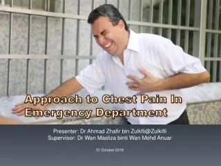
Approach to Chest Pain In Emergency Department Presenter: Dr Ahmad Zhafir bin Zulkfli@Zulkifli Supervisor: Dr Wan Maslizabinti Wan MohdAnuar 31 October 2018
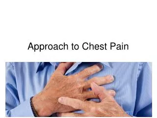
Introduction • Chest pain accounts for a large number of ED visits • Patients present with a spectrum of signs and symptoms reflecting the many potential etiologies of chest pain • Diseases of the heart, aorta, lungs, esophagus, stomach, mediastinum, pleura, and abdominal viscera may all cause chest discomfort • Clinicians in the ED focus on the immediate recognition and exclusion of life-threatening causes of chest pain • Patients with life threatening etiologies for chest pain may appear deceptively well, manifesting neither vital sign nor physical examination abnormalities
Differential • Cardiac • MI • Pericarditis • Myocarditis • Pulmonary • PE • Asthma/COPD • Pleura • Pleuritis • Pneumothorax • Aorta • Dissection • Chest wall • Costochondiritis/musculoskeletal pain • Esophagus • Esophageal Spasm • Esophagitis • Esophageal Rupture/Perforation • GERD • Panic attack
Characterized as discomfort/pressure rather than pain Time duration >2 mins Provoked by activity/exercise Radiation (i.e. arms, jaw) Does not change with respiration/position Associated with diaphoresis/nausea Relieved by rest/nitroglycerin Typical vs. Atypical Chest Pain Typical Atypical Pain that can be localized with one finger Constant pain lasting for days Fleeting pains lasting for a few seconds Pain reproduced by movement/palpation
Typical vs. Atypical Chest Pain UpToDate
Typical vs. Atypical Chest Pain Cayley 2005
Life threatening conditions • Acute coronary syndrome • Acute aortic dissection • Pulmonary embolism • Tension pneumothorax • Pericardial tamponade • Mediastinitis
ACS – STEMI/NSTEMI/USA • Result from atherosclerotic plaque rupture and thrombus formation • DEGREE and DURATION determine whether the patient develops reversible myocardial ischemia without injury (unstable angina) or myocardial ischemia with injury (myocardial infarction)
Pulmonary embolism • Estimated incidence over 1 in 1000 patients, but diagnosis is often missed • Occurs when a dislodged venous clot migrates through the right side of the heart and lodged at the branch point of the pulmonary arteries • Results in pulmonary hypertension, right ventricular dysfunction, poor gas exchange, and ultimately parenchymal infarction • Mortality rates vary widely based upon comorbid conditions and the size of the embolus • Early diagnosis and treatment reduce mortality for large hemodynamically unstable pulmonary emboli
Pneumothorax • Occurs following trauma, pulmonary procedures or spontaneously • Primary vs secondary pneumothorax • Regardless of etiology, the accumulation of air in the pleural space can lead to tension pneumothorax causing rapid clinical deterioration and death if unrecognized
Acute aortic dissection • Incidence of aortic dissection is estimated at 3 per 100,000 patients per year • Elderly, hypertensive patient younger patient in connective tissue disease • Results from tear in the inner layer of the aortic wall allowing blood to track between the intima and media
History • Onset of pain (eg, abrupt or gradual) • Provocation/Palliation (which activities provoke pain; which alleviate pain) • Quality of pain (eg, sharp, squeezing, pleuritic) • Radiation (eg, shoulder, jaw, back) • Site of pain (eg, substernal, chest wall, back, diffuse, localized) • Timing (eg, constant or episodic, duration of episodes, when pain began) • Associated symptoms: diaphoresis, nausea, and vomiting • Risk factors • Elderly >> atypical presentation!
Physical Examination • Most often the physical examination is not helpful in distinguishing patients with acute coronary syndromes (ACS) from those with noncardiac chest pain • Chest pain associated with focal wheezing or asymmetric extremity swelling raises concern for pulmonary embolus (PE) • Unilateral decreased breath sounds may be noted with pneumothorax • The presence of rales is associated with left ventricular dysfunction and left-sided heart failure • Pericardial friction rub in patients with pericarditis
Investigations • 12 leads ECG – AHA suggestion ECG obtained and interpreted within 10 minutes of patient presentation at ED • Serial ECG - increase the sensitivity for detecting ACS • Troponin I and T elevations within 3 hours, peak at 12 hours, and remain elevated for 7 to 10 days • FBC • CXR • Bedside ultrasound
Management • Evaluation of the chest pain patient in ED begins with assessment and stabilization of the airway, breathing, and circulation • Life-threatening problems are treated immediately, without delay for confirmatory testing • Patient is placed on a cardiac monitor and given supplemental oxygen if necessary while intravenous access is established • Obtain bloods, ECG and CXR
Management • Patients with ST elevation myocardial infarction (STEMI) require emergent revascularization via percutaneous intervention or fibrinolysis • Emergent treatment for a suspected aortic dissection involves blood pressure and heart rate control to reduce shearing forces and intensity of pulsatile cardiac flow • Tension pneumothorax is treated with immediate tube thoracostomy or immediate needle thoracostomyfollowed by tube thoracostomy • Patients with stable angina do not require inpatient evaluation
Case Discussion • 64 year old Malay male with underlying DM, HPT presents to ED HoSHAS with 5 hours of left sided chest discomfort associated with SOB, nausea and profuse sweating. Gradual onset while chopping trees in his estate. Partially improved with rest. • On examination:alert, conscious, pink, not tachypnic, warm peripheries, regular pulse volume, CRT <2S, profuse sweating • T 37.5ºC, BP 160/95mmHg, RR16bpm, PR 100bpm, spO2 98% under room air • Lungs clear, equal air entry • CVS s1s2 no murmur • Abdomen soft non tender • No calf tenderness, no pedal edema
Diagnosis: • If primary PCI cannot be performed, then fibrinolytic therapy should be administered with a DNT of less than 30 minutes
References • Cayley, W.E. Diagnosing the cause of chest pain. (2005). American Family Physician, Vol 72 (10), 2012-21 • Malaysian Clinical Practice Guideline on STEMI 3rd edition (2014), MoH • Evaluation of chest pain in the emergency department. UpToDate • Approach to patients with chest pain: differential diagnoses, evaluation and management, ECGWaves.com