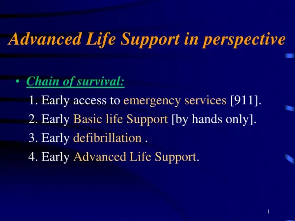
Advanced Life Support in perspective
Advanced Life Support in perspective. Chain of survival: 1. Early access to emergency services [911]. 2. Early Basic life Support [by hands only]. 3. Early defibrillation . 4. Early Advanced Life Support. Causes & prevention of Cardio respiratory arrest.

Advanced Life Support in perspective
E N D
Presentation Transcript
Advanced Life Support in perspective • Chain of survival: 1. Early access to emergency services [911]. 2. Early Basic life Support [by hands only]. 3. Early defibrillation . 4. Early Advanced Life Support.
Causes & prevention of Cardio respiratory arrest • Definition:A respiratory arrest is when breathing stops (apnea). A cardiac arrest is when the heart stops contracting & pumping blood. • Causes: 1. Airway problems. 2. Breathing problems. 3. Cardiovascular problems.
Airway Obstruction • Complete airway obstruction will rapidly result in cardiac arrest. • Partial airway obstruction may lead to cerebral or pulmonary edema , hypoxic brain damage as well as cardiac arrest . • Causes of airway obstruction [ blood, vomitus , F.B., direct throat / face trauma , CNSdepression, epiglottitis , epileptic fit, bronchialsecretions , mucosal edema, laryngeospasm , bronchospasm].
Cardiac Abnormalities • Primary causes [ventricular fibrillation]: 1. Ischemia. 2. M.I. 3. Drugs [digoxin , quinidine , phenothiazide , tricyclic antidepressant]. 4. Alcohol abuse. 5. Acidosis . 6. Abnormal electrolytes conc.[Ca, Mg & K].
Secondary causes of cardiac abnormalities: 1.asphyxia. 2. Apnea. 3. Acute sever blood loss. 4. Acute pulmonary edema. 5. Suffocation. 6. Hypoxemia , anemia , hypothermia , end-stage septic shock are having longer heart effect.
Prevention: 1. History, examination & investigation when needed. 2. Breathing problems is pre cardio respiratory arrest clinical abnormalities. 3. Hypotension , confusion , restlessness lethargy & L.O.C. should be considered . 4. Metabolic abnormalities particularly acidosis. 5. Consider ICU admission in your plan.
Ventilation • Face mask: [ 45 - 50% if more than 6 L/m ]. • Nasal Cannulae: [ 30 - 35% on 3 L/m]. • Ventorie: [ 24 – 90% ]. • Non re-breathing mask: [ 90% ]. • Laryngeal mask airway: [100% ]. • Endo tracheal tube: [100% ]. • Needle cricothyroidotomy: [full neck extension , feel the cricoid & prick 0.5 cm below it ].
Cardiac Monitoring & rhythm Recognition • Remember: Treat the patient not the ECG. • A normal HR is defined as 60 –100 b/m , a rate below 60 is known as bradycardia & a rate of 100 is known as tachycardia. • Rhythms causing cardiac arrest: 1. Supra-ventricular tachycardia [ above bundle of His bifurcation ]. 2. Ventricular tachycardia [distal to bifurcation].
Supra-ventricular tachycardia: 1. Atrial fibrillation: [absent P wave & normal QRS complex]. 2.Atrial flutter: [there is P wave but saw tooth in appearance & rate more than 200/m (250-300/m) with regular QRS complex]. 3.supra-ventricular tachycardia: [ you might find P wave or not , because it might start from A/V node ].
Ventricular tachycardia: 1.wide QRS complex. 2. rare more than 100/m. 3. may sustain for morethan 30 seconds (take it seriously). But if it was for less than 30 seconds it might be d.t. lytes imbalance or hypoxia. • Ventricular Fibrillation : 1. no pulse. 2. ECG show absent QRS & T wave & replaced by cont., very rapid, bizarre, irregular appearance of apparently random frequency & amplitude.
Drugs & Their delivery • Priority in drug delivery : 1. central line [30 seconds]. 2. Peripheral line [5 minutes]. 3. E.T. Tube [but we double or triple the IV dose]. 4. Intra Cardiac[ not used any more]: a) technically difficult. b) while doing the procedure CPR should stopped. c) high rate of complications: 1.coronary laceration. 2.intra mural injections. 3.pneumothorax.
Defibrillation • We paralyze the heart, to let S. A. Node to start working again . • The delay in DC >>>the sever the arrhythmia >>> less favorable prognosis & less responsive to treatment. • Types: 1. Synchronized Cardio-version. 2. A synchronized Cardio-version.
1. Synchronized Cardio-version: if is used to convert Atrial or ventricular tach., it is important that the shock is synchronized to occur with the R wave of the ECG rather than with the T wave. 2. A synchronized Cardio-version: it will shock at any ECG phase ,& it can cause ventricular fibrillation. • Mechanism of action: 1. Monophasic: receive single burst, 1 pad to another & don’t come back.
2. Biphasic : less Jules (electric shock waves move from 1 pad to the other then go in reverse direction). Types of Biphasic Defibrillator: 1. Manual (which we are using). 2. Shock Advisor (for non-expert people),with big electrodes they can read the rhythm then talk or write the order to be done. 3. Automated External (you just connect it to the patient & it will work & calculate the electric wave by it self & when to give it).
Position: 1. Right of the upper sternum below the clavicle 2. left 5th inter-costal space ant. Axillary's line. • Technique: 1. apply pressure to the paddle [10kg] to decrease thoracic impedance (the distance by pr. The fat). 2. keep the defibrillator paddles at least 12.5 cm from the pace maker if there is. 3. Keep oxygen flow away from from paddle (not to kill the patient by burning instead of arrest) 4. Don’t remove the paddle until 3 DC shock performed.
Treatment of Algorithms • During CPR: [If not already] 1. Check electrode/paddle position & contact. 2. Attempt/verify airway ,oxygen & IV access. 3. Give adrenaline every 3 minutes (cycle). 4. Consider : a. Anti-arrhythmic. b. Atropine.
[Correct Reversible causes (4 H’s & 4T’s)] 1. Hypoxia. 2. Hypovolemia. 3. Hypo/Hyperkalemia & metabolic disorders. 4. Hypothermia. 5. Tensionpneumothorax. 6. Tamponade. 7. Toxic/Therapeuticdisturbances. 8. Thrombo-embolic/mechanical obstruction.
Management of VF/pulse-less VT • In each CPR cycle we provide: • 1mg adrenaline IV. • 3 DC shocks (200, 200, & 360 joules). • 1 minute CPR. • Then after 1 min. CPR 3 DC shocks (each 360 joules)+ 1 minute CPR & adrenaline.
Algorithm for management of non VF/VT rhythms • In case of a systole there is no rule of DC shock unless fine VF. • 1mg adrenaline + 3mg atropine.(USA) • 3mg atropine but 0.5mg every 3 min & total of 3mg + 1mg adrenaline in each cycle. • Post DC shock heart can enter into a systole for 15 sec. Then return to normal.
Cardiac Arrest in special Circumstances • Hypothermia. • Near drowning. • Pregnancy. • Poisoning. • Electrocution. • Anaphylaxis. • Acute severe asthma.
Hypothermia • Hypothermia exist when the body core temp. falls below 35C. • (A&B) with high conc. Warm O2. • (C) palpate a major artery for a minimum of 1 minute before concluding that there is no C.O.P. • As body temp. falls sinus bradycardia A.F. V.F. finally a systole. When core temp < 30C ; VF will not respond to cardioversion or drugs. Arrhythmias other than VF tend to revert spontaneously as the core temp rises [in open heart surgery when we rise temp 33C the heart rate pick up systole & sinus rhythm]. • Rewarming: 1) Remove cold wet clothing ASAP & cover with blankets. 2) Warm bathes (40C).
Severe hypothermia (<28C) [maintain in ICU for 24 hrs]: 1) Ventilate with warm humidified O2. 2) I.V. warm Fluids (40C). 3) Gastric, peritoneal, or pleural lavage with warm fluids (@40C). 4) Heated blankets. 5) Blood rewarming by haemodialysis or cardiopulmonary bypass. N.B: - U.O.P. increase with hypothermia - Hypothermia promotes the transfer of fluid from the circulation into the tissues. - Warm slowly (1 degree/30 minutes).
Near Drowning • Associated with hypothermia & beadycardia. • Defined as asphyxiation in fluid (water). • Respiratory arrest 1ry event & cardiac arrest is 2ry event. • BLS & ALS shouldn’t be less than 45 min. • Placed horizontally & head down ( to prevent aspiration & regurgitation). • In 10% of cases no fluid is aspirated (dry drowning due to spasm). • Hospitalization is needed for: - Secondary pulmonary edema. - ARDS (aspirated fluid). • Patient can be discharged after 6 hrs if clinically, ABG, CXR normal.
Pregnancy • Two people to resuscitate. • Causes of maternal cardiac arrest: - Hemorrhage. - Pulmonary embolism. - Amniotic fluid embolism. - Placental abruption. - Eclampcia. • After 5 minutes of unsuccessful in-hospital resuscitation, emergency C/S is indicated to save the fetus (in the 3rd trimester of pregnancy).
Poisoning • A.B.C. & NO mouth to mouth breathing. • Increase risk of pulmonary edema, aspiration, early intubation is recommended in thermal injury & airway burns. • Arrhythmias commonly results from the ingestion of drugs with negative inotropic action (treatment is with positive inotropic drugs i.e. adrenaline & dobutamine).
Antidote: - OpiodXNaloxone 1.2mg - BradyarrhythmiaXatropine 2mg or isoprenaline 10-100ug/min. - B.blockersXglucagon 5mg IV. - Organophosphate insecticidesX high-dose atropine. - CyanidesXdicobalt edetate. - Digoxin toxicityXdigoxin specific FAB. • Pass NGT & lavage stomach from ingested toxins & give activated charcoal.
Electrocution • The severity of injury depends on the area & the magnitude & the path of the current. • Electricity tends to pass along muscles, nerves & vessels. It may therefore paralyze the respiratory muscles or disturb the myocardium, leading to respiratory or cardiac arrest (V. Fibrillation, immediate asystole, extra pace maker). • Electrocution is like a bullet goes in & out, but if it remains in it will settle at the heart. • Those who have survived an electric shock should be monitored in hospital if they have suffered (L.O.C, cardiac arrest, ECG abnormalities, contact injury)
Anaphylaxis • Due to (insect bite, food, blood products & drugs) IgE Anti bodies histamine release increase vascular permeability & peripheral V.D.( decrease V.R. & C.O.P.) sudden collapse & death. • Anaphylactoid reaction (there is no IgE mediators & no previous sensitization). • Resuscitation with: 1) 100% oxygen. 2) Adrenaline (if stridor, wheeze or respiratory distress) 0.5cc 1/1000 I.M. & repeat Q5 minutes if no clinical improvement is clear. 3) CPR or ALS. 4) Antihistamines. 5) Hydrocortisone. 6) IV Colloids.
Acute Severe Asthma • Normal or low PCO2. • Resuscitated with: 1) ABGs. 2) Intubation. 3) Exclude pneumothorax & consider open cardiac message. • Resist arrhythmias in case of metabolic disorders.
Peri-arrest • Arrhythmias complications of M.I. & in certain circumstances may also precede ventricular fibrillation, named : 1. Bradycardia. 2. Broad complex tachycardia (90% ventricular in origin). 3. Narrow complex tachycardia (90%atrial in origin). • Principle of treatment: 1. How is the patient ? 2. What is the arrhythmia ? • Three options available in the immediate treatment of arrhythmias: 1. Cardio-version. 2. Anti-arrhythmic & other drugs. 3. Cardiac pacing.
Bradycardia: blood pressure < 90 mmHg. • Broad complex tachycardia: 1) Pulse sedation synchronize DC shock… 2) Pulseless no C.O.P. VF/VT. • Narrow complex tachycardia: 1)AF > 130 b/min ask help BP < 90 mmHg synchronized DC shock. 2) Vagal maneuver adenosine ask help BP <90 mmHg synchronized DC shock.
Cardiac Pacing • SAN(60 - 70 beats/minute). • AVN(40 -50 beats/minute) [narrow QRS]. • His/Purkinje fibers(30 beats/minute) [wide QRS]. N.B. In open heart surgery the pace maker should be 100 beats/minute to over come SAN.
Artificial pacemakers classification: • Non-invasive: • Percussion pacing (decrease HR decrease COP). • Transcutaneous pacing (stickers). • Invasive: • Temporary transvenous pacing (central line placed in Rt. ventricle). • Permanent implanted pacing (catheter with patery). • Implantable cardioverter defibrillators.
patient is stable Thank you