Download

1 / 60
650 likes | 1.21k Vues
Lupus Erythematosus. Mohammed Al-Haddab, MD, FRCPC Assistant Professor & Consultant Dermasurgeon. Dept. of Dermatology. College of Medicine. King Saud University. Objectives . At the conclusion of these lectures the student will be able to:

E N D
Lupus Erythematosus Mohammed Al-Haddab, MD, FRCPC Assistant Professor & Consultant Dermasurgeon. Dept. of Dermatology. College of Medicine. King Saud University
Objectives At the conclusion of these lectures the student will be able to: • differentiate between the various types of Lupus • recognize how Lupus affects the various systems of the body • identify all of the current treatment options available for Lupus • recognize the psychosocial effects that Lupus has on the patient and their family
Objectives • To learn how to diagnose and investigate dermatomyositis. • How to manage dermatomyositis. • To learn the presentation of morphea and systemic sclerosis and ways to manage them. • To recognize other diseases like PG, lichen sclerosis and how to manage them. • This lecture is not meant to be inclusive of all the information about these diseases but to highlight important aspects in their diagnosis and management.
Lupus Erythematosus LE is as an autoimmune diseases associated with antibodies directed against components of cell nuclei. Lupus may affect any tissue, skin, kidneys, CNS, lungs and others. Discoid lupus erythematosus Subacute lupus erythematosus Neonatal lupus erythematosus Lupus tumidus Lupus profundus Chilblain lupus erythematosus Drug-induced lupus erythematosus Systemic Lupus Erythematosus
Discoid Lupus Erythematosus It is the commonest form of cutaneous lupus usually presents as red scaly patches or plaques that leave dyspigmentation and scarring mostly Hypopigmented or depigmented scars. It may be localized or widespread. Usually affects the cheeks, nose and ears, but sometimes involves the upper back, V of neck, and backs of hands. Involvement of hair follicles will lead to scarring alopecia.
Subacute lupus erythematosus a non-itchy dry rash appears on the upper back and chest, often following sun exposure. Subacute LE does not scar. And systemic involvement is not usually severe. Annular or polycyclic (ring-shaped) or as papulosquamous (scaly patches and plaques)
Neonatal lupus erythematosus Newborn babies born to mothers with subacute LE may develop annular rash, known as neonatal LE that resolve spontaneously. The neonates could be at risk of complete heart block.
Lupus Tumidus a dermal form of lupus. The rash is characteristically photosensitive, so it affects sun-exposed sites. It presents with red, swollen, urticaria-like bumps and patches or swelling.
Lupus Profundus lupus affecting the fat underlying skin lupus panniculitis. it may develop at any age, including children. The face is the most common area to be affected. Inflammation of the fat results in firm deep nodules for some months. The end result is deep scars on fat layer or lipodystrophy.
Chilblain Lupus Erythematosus Chilblains (or pernio) are itchy and/or tender red or purple bumps that usually come on from cold exposure but can sometimes be precipitated by sun exposure or smoking. They are considered to be a form of skin vasculitis (blood vessel inflammation). They can occur in people with lupus or in otherwise healthy people, especially children and the elderly. Usually they have no circulating antibodies. And the main treatment is to avoid precipitating factors.
Drug-Induced Lupus Erythematosus Drug induced lupus does not usually affect the skin. The most frequent drugs are: Hydralazine , Carbamazepine , Lithium , Phenytoin , Sulphonamides , Minocycline.
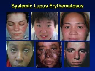
Systemic Lupus Erythematosus Only a few patients with cutaneous LE also have SLE. The most common presentation is with a malar eruption or butterfly. Other skin changes in SLE are photosensitivity, mouth ulcers, and diffuse hair loss. SLE may also affect joints, kidneys, lungs, heart, liver, brain, blood vessels and blood cells
Investigations SLE is always with positive ANA. antiRo/La antibodies, is nearly always present in patients with subacute LE. Leucopenia tends to be more pronounced in patients with systemic LE Skin biopsy may be diagnostic especially in discoid lupus erythematosus. Direct immunofluorescence tests may show positive antibody deposition along the basement membrane (lupus band test).
Treatment of Cutaneous Lupus Erythematosus The aim of treatment for cutaneous LE is to alleviate symptoms and to prevent scarring. Smoking cessation will help Raynaud's phenomena and chilblain lupus . Sun protection. Potent topical steroids, Intra lesional steroids. Oral antimalarial drugs. Oral steroids. Methotrexate, azathioprin, mycophenolatemofetil, cyclosporine, cyclophosphamide, IVIG, and Rituximab.
Dermatomyositis An uncommon inflammatory disease affects adults between 40-60 (females mainly) and children 5-15. Skin changes. A violet-colored or dusky red rash on face and eyelids and on areas around nails, knuckles, elbows, knees, chest and back. The rash, which can be patchy with bluish-purple discolorations, is often the first sign of dermatomyositis. Muscle weakness. Progressive proximal muscle weakness involves the hips, thighs, shoulders, upper arms and neck. The weakness is symmetrical and more in the extensor muscles.
Dermatomyositis Other signs and symptoms include: Photosensitivity Raynaud's phenomenon Dysphagia, gastrointestinal ulcers Muscle pain or tenderness Fatigue, fever and weight loss Calcinosis cutis especially in children Interstitial lung disease.
Dermatomyositis It can be associated with: Other connective tissue diseases such as lupus, rheumatoid arthritis, scleroderma and Sjogren's syndrome. Cancer, Especially in older patients, particularly of the cervix, lungs, pancreas, breasts, ovaries and gastrointestinal tract. Cancer could precede, coincide or follow the diagnosis of DM.
Investigations • Magnetic resonance imaging (MRI). • Electromyography. • Muscle biopsy. • Blood tests: creatine kinase (CK) and aldolase. Increased CK and aldolase levels can indicate muscle damage and CK is useful to monitor the treatment of DM. • autoantibodies • Skin biopsy is suggestive but not diagnostic that shows interface dermatitis.
Treatment Oral steroids are the mainstay treatment. Steroid sparing agents are: Methotrexate, azathioprin, mycophenolatemofetil, cyclosporine, cyclophosphamide, IVIG, and Rituximab. Topical steroids and antimalarial medications are used to improve the cutaneous rashes. Physiotherapy to improve strength and flexibility of the muscles. Surgical excision or Co2 laser could be utilized to remove tender calcium deposits .
Scleroderma A group of rare diseases that involve the hardening and tightening of the skin and connective tissues Scleroderma affects women more often than men and most commonly occurs between the ages of 30 and 50.
Morphea a rare skin condition that causes oval reddish or purplish patches and plaques on the skin. Sometimes in linear distribution on face and extremities. It subsides on its own over time leaving dyspigmentation and scars. diagnosed on the base of its morphology and confirmed by skin biopsy which usually shows thickening of collagen bundles and loss of skin appendages like sweat glands and hair follicles. Morphea has no known cure. Treatment of morphea focuses on controlling signs and symptoms and slowing spread. Topical and intralesional steroids , phototherapy, systemic steroids, azathioprine, methotrexate, and cyclosporine might be used in severe cases. Physical therapy could be of help if the involvement is close to joints and cause contracture and difficulty movement.
CREST Syndrome Is a limited form of systemic sclerosis in which there is Calcinosis, Raynaud's phenomenon, Esophageal involvement, Sclerodactyly and Telangiectases. Anticentromere antibodies are characteristic for this syndrome.
Systemic Sclerosis An autoimmune multisystem disease that results in fibrosis and vascular abnormalities in association with autoimmune changes. usually starts between 30-40 years in women who are more affected and later in men. Pathophysiology may involve some injury to the endothelial cells and this results in excessive activation of the dermal connective tissue cells, the fibroblasts. Usually presents with Raynaud's phenomena, Thickening of the skin of the fingers, then atrophy and sclerosis. The fingers become spindle-shaped (sclerodactyly) from resorption of the fingertips. Fragile nails become smaller with ragged cuticles The tight shiny skin may affect most parts of the body, including the face, resulting in loss of expression and difficulty opening the mouth properly.
Systemic Sclerosis telangiectasia appear on the fingers, palms, face, lips, and chest. Ulcers may follow minor injuries over the joints, or on the tips of fingers and toes. Ulceration can lead to dry gangrene and eventual loss of the tips of the fingers Joint contractures. Patients will be bed ridden with time. Esophageal reflux and dysphagia. Lung and heart involvement may manifest as shortness of breath, high blood pressure, chest pain, pleurisy, pneumothorax, pericarditis arrhythmias, general heart enlargement and heart failure. Progressive kidney disease resulting in proteinuria, high blood pressure and eventually renal failure.
Systemic Sclerosis Diagnosis is made based on clinical features and presentation. Skin biopsy will show skin atrophy with preservation of skin appendages. ANA is usually positive. Anti topoisomerase I (Scl 70) is characteristic for it especially in severe cases.