Download
1 / 70
710 likes | 1.43k Vues
Chapter 27: The Head, Face, Eyes, Ears, Nose and Throat. Prevention of Injuries to the Head, Face, Eyes, Ears, Nose and Throat. Head and face injuries are prevalent in sport, particularly in collision and contact sports

E N D
Prevention of Injuries to the Head, Face, Eyes, Ears, Nose and Throat • Head and face injuries are prevalent in sport, particularly in collision and contact sports • Education and protective equipment are critical in preventing injuries to the head and face • Head trauma results in more fatalities than other sports injury • Morbidity and mortality associated w/ brain injury have been labeled the silent epidemic
Assessment of Head Injuries • Brain injuries occur as a result of a direct blow, or sudden snapping of the head forward, backward, or rotating to the side • May or may not result in loss of consciousness, disorientation or amnesia; motor coordination or balance deficits and cognitive deficits • May present as life-threatening injury or cervical injury (if unconscious)
History • Determine loss of consciousness and amnesia • Additional questions (response will depend on level of consciousness) • Do you know where you are and what happened? • Can you remember who we played last week? (retrograde amnesia) • Can you remember walking off the field (antegrade amnesia) • Does your head hurt? • Do you have pain in your neck? • Can you move your hands and feet?
Observation • Is the athlete disoriented and unable to tell where he/she is, what time it is, what date it is and who the opponent is? • Is there a blank or vacant stare? Can the athlete keep their eyes open? • Is there slurred speech or incoherent speech? • Are there delayed verbal and motor responses? • Gross disturbances to coordination?
Inability to focus attention and is the athlete easily distracted? • Memory deficit? • Does the athlete have normal cognitive function? • Normal emotional response? • How long was the athlete’s affect abnormal? • Is there any swelling or bleeding from the scalp? • Is there cerebrospinal fluid in the ear canal?
Palpation • Neck and skull for point tenderness and deformity • Special Tests • Neurologic exam (pg 35-352) • Assess cerebral testing, cranial nerve testing, cerebellar testing, sensory and reflex testing • Eye function • Pupils equal and reactive to light (PEARL) • Dilated or irregular pupils • Ability of pupils to accommodate to light variance • Eye tracking - smooth or unstable (nystagmus, which may indicate cerebral involvement) • Blurred vision
Balance Tests • Romberg Test • Assess static balance - determine individual’s ability to stand and remain motionless • Multiple variations (primarily foot position) • feet together, single non-dominate leg, tandem (heel/toe) • Perform both on solid ground and on balance pad • Balance Error Scoring System (BESS) • Quantifiable clinical battery of test that utilizes different stances on both firm and foam surface • Errors are tabulated when the athlete opens their eyes, takes hands off hips, steps/stumbles or falls. • 2 trials each surface x 20 secs ea. • >10 total errors = failure • Coordination tests • Finger to nose, heel-to-toe walking • Inability to perform tests may indicate injury to the cerebellum
BESS 2 x 20 secs ea. Score using BESS worksheet Count number of errors per 20 sec session > 10 point errors = failure
Cognitive Tests • Used to establish impact of head trauma on cognitive function and to obtain objective measures to assess patient status and improvement • On or off-field assessment • Serial 7’s, months in reverse order, counting backwards • Tests of recent memory (score of contest, breakfast game, 3 word recall) • Neuropsychological Assessments • Standardized Assessment of Concussion (SAC) provides immediate objective data concerning presence and severity of neurocognitive impairment • Used to assess orientation, immediate memory recall, concentration, and delayed recall on and off the field
Neuropsychological Assessment (continued) • Other assessment tools have been designed to assess short term memory, working memory, attention, concentration, visual space capacity, verbal learning, information processing speed and reaction time • Computerized neuropsychological testing programs have been developed • IMPACT computer testing • Computer based base-line neurocognitive assessment test for concussion management • Univ of Pittsburgh medical center • Standard of care expecation
Complete the following Head Injury Assessment Tests in lab • Eye Function • PEARL test • Eye Tracking • Eye Chart Testing • Balance Tests • Romberg • BESS – use grading chart provided in note • Coordination Tests • Cognitive Tests • Neuropsychological Tests • SAC – use grading chart provided in notes • IMPACT computer – print finished test
Recognition and Management of Specific Head Injuries • Skull Fracture • Etiology • Most common cause is blunt trauma • Signs and Symptoms • Severe headache and nausea • Palpation may reveal defect in skull • May be blood in the middle ear, ear canal, nose, ecchymosis around the eyes (raccoon eyes) or behind the ear (Battle’s sign) • Cerebrospinal fluid may also appear in ear and nose • Management • Immediate hospitalization and referral to neurosurgeon
Cerebral Concussions (Mild Head Injuries) • Characterized by immediate and transient post-traumatic impairment of neural function • Etiology • Result of direct blow, acceleration/deceleration forces producing shaking of the brain • Signs and Symptoms • Brief periods of diminished consciousness or unconsciousness that lasts seconds or minutes • Glasgow Coma score of 13 -15 • Post-traumatic amnesia lasting <24 hours • No signs of focal injury (subdural or epidural hematoma) • Negative CT or MRI imaging studies
Management • The decision to return an athlete to competition following a brain injury is a difficult one that takes a great deal of consideration • If any loss of consciousness occurs the ATC must remove the athlete from competition • With any loss of consciousness (LOC) a cervical spine injury should be assumed • Objective measures (BESS and SAC) should be used to determine readiness to play • A number of guidelines have been established to in an effort to aid clinicians in their decisions • Return to normal baseline requires approximately 3-5 days
Management (continued) • All post-concussive symptoms should be resolved prior to returning to play -- any return to play should be gradual • Recurrent concussions can produce cumulative traumatic injury to the brain • Following an initial concussion the chances of a second episode are 3-6 times greater
Postconcussion Syndrome • Etiology • Condition which occurs following a concussion • May be associated w/ those MHI’s that don’t involve a LOC or in cases of severe concussions • Signs and Symptoms • Athlete complains of a range of postconcussion problems • Persistent headaches, impaired memory, lack of concentration, anxiety and irritability, giddiness, fatigue, depression, visual disturbances • May begin immediately following injury and may last for weeks to months • Management • ATC should treat symptoms to greatest extent possible • Return athlete to play when all signs and symptoms have fully resolved
Second Impact Syndrome • Etiology • Result of rapid swelling and herniation of brain after a second head injury before symptoms of the initial injury have resolved • Second impact may be relatively minimal and not involve contact w/ the cranium • Impact disrupts the brain’s blood autoregulatory system leading to swelling, increasing intracranial pressure • Signs and Symptoms • Often athlete does not LOC and may looked stunned • W/in 15 seconds to several minutes of injury athlete’s condition degrades rapidly • Dilated pupils, loss of eye movement, LOC leading to coma, and respiratory failure
Second Impact Syndrome (continued) • Management • Life-threatening injury that must be addressed w/in 5 minutes w/ life saving measures performed at an emergency facility • Best management is prevention from the ATC’s perspective
Cerebral Contusion • Etiology • Focal injury to the brain that involves small hemorrhages or intracranial bleeding w/in the cortex, stem or cerebellum • Generally occurs when head strikes a stationary object • Signs and Symptoms • Severity will vary greatly based on the extent of the injury • Will likely experience a LOC followed by a very talkative state • Normal neurological exam; presenting w/ headache, dizziness and nausea • Management • Hospitalization w/ CT and MRI • Treatment will vary according to status of the athlete • Return to play occurs when athlete is asymptomatic and CT is normal
Epidural Hematoma • Etiology • Blow to head or skull fracture which tear meningeal arteries • Blood pressure, blood accumulation and creation of hematoma occur rapidly (minutes to hours) • Signs and Symptoms • LOC followed by period of lucidity, showing few signs and symptoms of serious head injury • Gradual progression of S&S • Head pains, dizziness, nausea, dilation of one pupil (same side as injury), deterioration of consciousness, neck rigidity, depression of pulse and respiration, and convulsion • Management • Requires urgent neurosurgical care; CT may be necessary for diagnosis • Must relieve pressure to avoid disability or death
Subdural Hematoma • Etiology • Result of acceleration/deceleration forces that tear vessels that bridge dura mater and brain • Venous bleeding (simple hematoma may result in little to no damage to cerebellum while more complicated bleed can damage cortex) • Signs and Symptoms • With a simple subdural hematoma LOC generally does not occur • Complicated subdural hematoma’s result in LOC, dilation of one pupil • Both will show signs of headache, dizziness, nausea or sleepiness • Management • Immediate medical attention • CT or MRI is necessary to determine extent of injury
Malignant Brain Edema Syndrome • Etiology • Occurs in young athletes w/in minutes to hours of a head injury • Caused by intracerebral clot resulting in diffuse brain swelling w/ little or no brain injury • Swelling is the result of hyperemia or vascular engorgement - results in increased pressure • Signs and Symptoms • Rapid neurologic deterioration that progresses coma and occasionally death • Management • Life-threatening condition requiring immediate attention at an emergency care facility
Migraine Headaches • Etiology • Disordered characterized by recurrent attacks of severe headache • Seen in those that have had repeated head trauma • Exact cause unknown (believed to be vascular) • Signs and Symptoms • Sudden onset w/ possible visual or gastrointestinal problems • Flashes of light, blindness (half field vision), paresthesia • Management • Prevention is key • Prescription medications have a high success rate
Scalp Injuries • Etiology • Blunt trauma or penetrating trauma tends to be the cause • Can occur in conjunction with serious head trauma • Signs and Symptoms • Athlete complains of blow to the head • Bleeding is often extensive (difficult to pinpoint exact site) • Management • Clean w/ antiseptic soap and water (remove debris) • Cut away hair if necessary to expose area • Apply firm pressure or astringent to reduce bleeding • Wounds larger than 1/2 inch in depth should be referred • Smaller wounds can be covered w/ protective covering and gauze (use extra adherent)
Recognition and Management of Specific Facial Injuries • Mandible Fractures • Etiology • Direct blow (generally fractures at frontal angle) • Signs and Symptoms • Deformity, loss of occlusion, pain with biting, bleeding around teeth, lower lip anesthesia • Management • Temporary immobilization w/ elastic wrap followed by reduction and fixation
Mandibular Dislocation • Etiology • Involves TMJ joint • MOI is generally a side blow to an open mouth • Signs and Symptoms • Dislocated jaw presents in locked-open position w/ ROM minimal along w/ poor occlusion • Management • Cold application, elastic wrap immobilization and reduction • Follow-up w/ soft diet, NSAID’s and analgesics w/ a gradual return to activity 7-10 days following acute period • Can be recurrent or result in malocclusion, or TMJ dysfunction
Tempromandibular Joint Dysfunction • Etiology • Disk condyle derangement (disk is positioned anteriorly) • Signs and Symptoms • Headaches, earaches, vertigo, inflammation, neck pain, muscle guarding and trigger points • Hyper- or hypomobility, muscle dysfunction, limited ROM, clicking and popping • Management • Treat with custom designed, removable mouth piece • Treat problem w/ either strengthening or stretching • If corrective measures fail, referral to a dentist will be necessary
Zygomatic complex (cheekbone) fracture • Etiology • MOI = direct blow • Signs and Symptoms • Deformity, or bony discrepancy, nosebleed, diplopia, and numbness in cheek • Management • Cold application to control edema and immediate referral to a physician • Healing will take 6-8 weeks and proper gear will be required upon return to play
Facial Lacerations • Etiology • Result of a direct impact, and indirect compressive force or contact w/ a sharp object • S&S • Pain, substantial bleeding, • Management • Apply pressure to control bleeding • Referral to a physician will be necessary for stitches
Prevention of Dental Injuries • When engaged in contact/collision sports mouth guards should be routinely worn • Greatly reduces the incidence of oral injuries • Practice good dental hygiene • Dental screenings should occur yearly • Cavity prevention • Prevention of abscess development, gingivitis, and periodontitis
Tooth Fractures • Etiology • Impact to the jaw, direct trauma • Signs and Symptoms • Uncomplicated fractures produce fragments w/out bleeding • Complicated fractures produce bleeding, w/ the tooth chamber being exposed w/ a great deal of pain • Root fractures are difficult to determine and require follow-up w/ X-ray
Tooth Fractures (continued) • Management • Uncomplicated and complicated crown fractures do not require immediate attention • Fractured pieces can be placed in a bag and and if not sensitive to air or cold, follow-up can wait for 24-48 hours • Bleeding can be controlled via gauze • Cosmetic reconstruction of tooth • In instances of root fractures, the athlete can continue to play but must follow-up immediately following competition • Tooth repositioning may be required, along with bracing and the use of mouthpieces in the future • Mandibular fractures and concussions must also be ruled out
Tooth Subluxation, Luxation and Avulsion • Etiology • Direct blow • Signs and Symptoms • Tooth may be slightly loosened, dislodged • When subluxed tooth may be loose w/in socket w/ little or no pain • With luxations, no fracture has occurred, however, there is displacement • W/ an avulsion, the tooth is completely knocked from the oral cavity • Management • For a subluxed tooth, referral should occur w/in the first 48 hours • With a luxated tooth, repositioning should be attempted along w/ immediate follow-up • Avulsed teeth should not be re-implanted except by a dentist (use a Save a Tooth Kit, milk or saline)
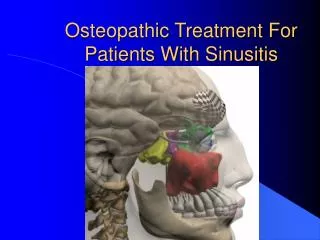
Nasal Injuries • Nasal Fractures and Chondral Separation • Etiology • Direct blow • Signs and Symptoms • Separation of frontal processes of maxilla, separation of lateral cartilage or combination • Profuse bleeding and hemorrhaging, immediate swelling and deformity
Management • Control bleeding and refer to a physician for X-ray,examination and reduction • Uncomplicated and simple fractures will pose little problem for the athlete’s quick return • Splinting may be necessary
Deviated Septum • Etiology • Compression or lateral trauma • Signs and Symptoms • Bleeding and in some instances a septal hematoma • Athlete will complain of nasal pain • Management • At the site of the hematoma, compression will be required (and if present, drained immediately) • Following drainage, a wick is inserted to allow for further drainage • Packing will be necessary to prevent a return of the hematoma • A neglected hematoma will result in formation of an abscess along with bone and cartilage loss and deformity
Nosebleed (epistaxis) • Etiology • Result of a direct blow, a sinus infection, high humidity, allergies, a foreign body or some other serious facial injury • Signs and Symptoms • Generally bleeding from the anterior aspect of the septum • Generally presents with minimal bleeding and resolves spontaneously • More severe bleeding may require more medical attention
Management • W/ acute bleeding, sit upright w/ a cold compress over the nose, pressure on the affect nostril and the ipsilateral carotid artery • Also gauze between the upper lip and gum - limits blood supply • If bleeding does not cease in 5 minutes, an astringent or styptic may need to be applied along with a gauze/cotton nose plug to encourage clotting • After bleeding has ceased, the athlete can return to play but should be reminded not to blow the nose under any circumstances for at least 2 hours after the initial insult
Recognition and Management of Specific Ear Injuries • Auricular Hematoma (Cauliflower Ear) • Etiology • Occurs either from compression or shear injury to the ear (single or repeated) • Causes subcutaneous bleeding
Auricular Hematoma (Cauliflower Ear) • Signs and Symptoms • Tearing of overlying tissue away from cartilage • Hemorrhaging and fluid accumulation • If unattended - coagulation, organization and fibrosis occurs • Appears as elevated, white, rounded nodular formation, that is firm and resembles cauliflower • Management • To prevent, wear proper ear protection • Cold application will minimize hemorrhaging • If swelling occurs, measures must be taken to prevent fluid solidification • Physician aspiration, packing, pressure
Rupture of the Tympanic Membrane • Etiology • Fall or slap to the unprotected ear or sudden underwater variation can result in a rupture • Signs and Symptoms • Complaint of loud pop, followed by pain in ear, nausea, vomiting, and dizziness • Hearing loss, visible rupture (seen through otoscope) • Management • Small to moderate perforations usually heal spontaneously in 1-2 weeks • Infection can occur and must be continually monitored