Download
1 / 35
920 likes | 3.98k Vues
Rehabilitation Program for Patellofemoral Pain Syndrome. Chris Pantellere Dr. Sterner April 2009. What is PFPS?.

E N D
Rehabilitation Program for Patellofemoral Pain Syndrome Chris Pantellere Dr. Sterner April 2009
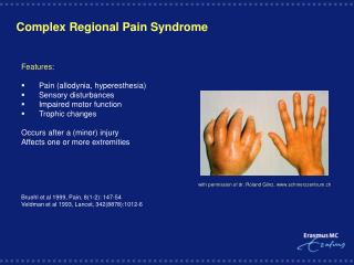
What is PFPS? • Defined as: a pathologic condition resultant of many different factors that produces retropatellar or prepatellar pain that is associated with the articulation between the femoral condyles and the undersurface of the patella • Can be a result of • Overactivity • Muscular imbalance • Malalignment of the lower extremity • Malalignment • Lower extremity malalignment • Patellar malalignment
Anatomy • Femur • Tibia • Patella
Musculature • Quadriceps • Iliotibial Band (ITB) • Adductor Magnus • Gracilis • Sartorious • Hamstrings • Gastrocnemius • Non-contractile components • Medial and lateral retinaculum
Biomechanics • The PF joint is placed under many different levels of stress at different ROMs. This is also dependent whether the individual is in a WB or NWB position. • Why is this important? • Because with out proper understanding of stresses placed on PF joint we may cause more damage/pain • Patellofemoral compressive forces generally increase with knee flexion and decrease with knee extension. Maximal patellofemoral compression 40-70 degrees of flexion • Dependent if the exercise is closed chain or open chain the PF joint will be under different compressive forces
Open vs. closed chain biomechanics • Open chain • PF compressive forces are greatest between 0o and 30o • Closed Chain • PF compression forces are greatest between 60o and 90o • Open and closed chain positions as well as the degree of flexion/extension can affect which muscles are actively being used…
Muscle Activity • Open Kinetic Chain exercises • More rectus femoris activity at angles less than 65o • Vastusmuslces more active in angles less than 45o • Closed kinetic chain exercises • More rectus femoris activity between 83-95o of flexion • Vastus muscles produced 50% more activity than rectus femoris • Greater than 55o the more vastus activity • Overall there is more Vastus muscle activity especially the vastusmedialis • 20% more than OKC
Patellar tracking Articulating surfaces of the patella with femur during different degrees of flexion/extension Degrees of motionArticulating surface 0 Degrees Fat pad which sits below patella 20 Degrees Inferior surface of patella 45 Degrees Medial Facet of patella 60-90 Degrees The greatest amount of compressive forces on the knee. The medial and lateral facets will articulate the most throughout the intercondylar notch. 135 Degrees Odd Facet
Etiology • Malalignment of lower extremity • Increased Q angle • GenuValgum • Femoral neck anteversion • Excessive pronation of the mid foot • Patella malalignment • Secondary to LE alignment • Patella alta, baja, squinting • Could be secondary to muscular tightness as well • Muscular imbalance • Over active vs. underactive • Asynchronous firing of VMO and VML • Overactivitiy • Lateral retinaculum tightness
Assessment • History • Insidious Onset • Diffuse Pain (possibly underneath the patella) • Buckling and instability ascending and descending stairs • Change in activity (training vol, surfaces, etc.) • As clinicians we must … • Full assessment of lower extremity to find the causes or contributing factors of PFPS. • Start distally work up to pelvis. • Check tightness, strength, weaknesses of musculature • Treat all the factors not just the most glaring.
Rehabilitation Protocol • Phase 1: 2-4 weeks • Phase 2: 2-4 weeks • Phase 3: 2-4 weeks • Phase 4: Functional testing
Phase 1 • Phase 1 of this rehabilitation program is primarily gauged at increasing ROM and general strengthening • Should focus on the areas of concern brought upon evaluation • Dependent on severity athlete may continue limited participation while rehabilitating. • Many cases we will allow activity but rehabilitate daily.
Phase 1: stretching • ITB** • Hamstrings • Quadriceps/hip flexors • Groin musculature
Phase 1: Joint mobilizations • Medial Patellar Glide most important • Perform with the knee in multiple positions (except full flexion) • Progress from 0o • Stretching the Lateral retinaculum • Begin with Grade 1 and 2 and progress to grades 3 and 4 • Grades I and II- pain reduction and relaxation (2-3 oscillations 1-2 min) • Grades III and IV- increase mobility (2-3 oscillations fro 20-60 sec) • Repeat 3-5 times
Phase 1: NMC training • Basics; begin shoes on eyes open on stable surface • Progress to shoes off • Then eyes closed • 30 seconds; record errors • T-band kicks • All should be performed with 30o flexion • Optimal co-contraction
Phase 1: Strengthening • Open Kinetic Chain Exercises- all 3sets 12 repetitions • Quad sets • Manual resisted knee extension • Straight leg raises • Hip flexion • Extension • Abd/add • Movements dependent on areas of weakness • Muncie Method • Towel squeeze • Isometric abd • Use light weights and gravity
Phase 1: Strengthening • Closed kinetic chain exercises- 3x12 • Terminal knee extension with theraband • Use biofeedback if available. • Leg presses- 45o • Squats- 45o • Stances can be changed by athlete • Can make more challenging • No difference in muscle activity!!! • Front/lateral step ups • Wall squats • 45o • Isometric Wall sits • 30 sec hold • Progress to 2 min
Phase 1: Cardiovascular training • Aquatics • Running in water • Swimming; freestyle • Eliptical • Bicycle • Be aware of ROMs that cause pain • Maintenance • requires 60% of VO2 max for 20-30 min a day • Sprint programs on bike
Phase 2 • Progressing to phase 2 when… • Inflammation significantly reduced • Pain only occurs past 90o of CKC • Significant strength gains in VMO and other quad musculature • Sustained contraction (biofeedback) • Increased ROM in hamstrings, quads, adductors • Dependent on where tightness is
Phase 2 • Key points… • Continue stretching (specifically ITB) • Continue Patellar mobilizations • Continue NMC- progress • Continue strengthening • Emphasize CKCE • Increase weight • Increase ROM of exercises • Continue Cardiovascular fitness
2 3 1 4 Phase 2: Neuromuscular control • Progress to unstable surface with no shoes • Progress to unstable surface with eyes closed • Sport specific activities while performing • Quadrant hops • Progression • Begin going clockwise/counter clockwise • 1,3,2,4 • Call out numbers • Eventually get to eyes closed • Add resistance with theraband • Can incorporate 180o rotations with hop. • Very versatile.
Phase 2: NMC continued • Lunges on an unstable surface- 3x12 • Straddled position; one leg on unstable surface • Hands on hips • 90o bend • Can do forward or backward lunges • Progress to eyes closed • Squats on bosu ball 3x12 • Can do on rounded surface or on flat platform on the bottom
Phase 2: strengthening • Closed kinetic chain exercises- 3x15 • Wall squats to 90o • Incorporate adduction squeeze with medicine ball • Wall sits @ 90o • Leg Press • Deeper ROMs • Barbell squats • 90o • Front lunges • Reverse lunges
Phase 2: Isokinetic strengthening • Concentric/eccentric protocol of quad musculature • Begin at lower velocities work up to more functional velocities. • Studies have shown that isokinetic strengthening of quad musculature for 8 weeks relieved the symptoms in 70% of patients with PFPS. Typical progressions: 90o per sec 150o per sec 225o per sec Can move to higher velocities to mimic more functional activites
Phase 2: functional activity • Running, cutting, jumping • Straight line jogging • Large figure 8’s • T drills • Sprint lateral shuffle • Agility ladder • Lateral 2 in 2 out • Forward 2 in 2 out • Lateral “ickey” shuffle • Scissor jumps
Phase 3 • Progressing to Phase 3 when… • Bilateral strength in VMO • Pain free ROM in CKCE and OKCE • Perform neuromuscular control activities w/ no errors
Phase 3 • Key points… • Continue stretching (specifically ITB) • Continue cardiovascular fitness • Move away from OCKE and more towards more dynamic CCKE • Progress weights and increase ROMs • Continue Dynamic NMC exercises with no errors • Patellar Mobilization • Increase Running and Cutting and agility drills • Begin Plyometric Activities
Phase 3: functional activities • Sprinting • Smaller Figure 8’s • T-Drill (sprint) • Four Corner Drill • Agility ladder drills • Use more dynamic drills • Modified Four Corner Drill • 4-3-2-1 • 1-2-4-3-1 • Sport Specific • Shuffle, back peddle, carioca
Phase 3: Plyometrics • Begin with the basics • As they become better and times improve progress to more dynamic forms of plyometrics • Side to side ankle hops (over barrier): 2x 16 • Squat with overhead medicine ball toss: 2x 25 • Single leg side to side ankle hops(over barrier): 2x 16 • Lateral step ups: 2x 20 • Squat jumps • Lunge jump (beginner)
Phase 3: Advanced Plyometrics • Depth jump with vertical jump: 2x10 • Depth jump with long jump: 2x10 • Single Leg Depth Jump: 2x10 • Depth Jump With Lateral Movement • Double leg barrier jump: • Emphasize soft landing • There and back (5 barriers) • Lunge jumps (advanced):2x 15 • Tuck jumps: 2x 15 • Standing Long Jump With Sprint: 2x10 • Standing Long Jump With Lateral Sprint: 2x 10 • Barrier hop with 180 degree turn: 2x 10
Phase 4: Sport specific training/return to play • Several different running, cutting, jumping, and landing activities • Place athlete in compromising positions • Modify for sport • Add barriers or obstacles to work with • Pain free • 2 to 3 Sets w/ 90 Seconds Rest • Should occur on the athlete’s competition field • Include some plyometrics with change of direction and sport specific movements (shuffling, backpedals, etc…)
Phase 4: Sport specific/RTP activities Forward two in two out x 5 single leg side to side jump over cone with turn and sprint Perform 10 squat jumps to sprint x end Start