Download
1 / 38
380 likes | 794 Vues
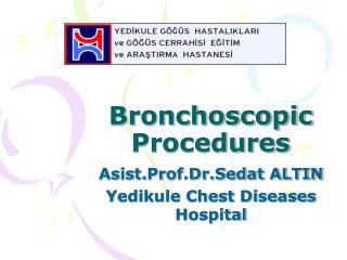
Virtual Bronchoscopic approach for combining 3D CT and endoscopic video. Anthony J. Sherbondy, 1 Atilla P. Kiraly, 1 Allen L. Austin, 1

E N D
Virtual Bronchoscopic approach for combining 3D CT and endoscopic video Anthony J. Sherbondy,1 Atilla P. Kiraly, 1 Allen L. Austin,1 James P. Helferty, 1 Shu-Yen Wan,1 Janice Z Turlington,1 Tao Yang,1 Chao Zhang,1 Eric A. Hoffman,2 Geoffrey McLennan,2 and William E. Higgins1,2 1Penn State University, University Park, PA 16802 2University of Iowa, Iowa City, IA 52246 SPIE Med. Imaging 2000, San Diego, CA, 12 February 2000
Virtual Endoscopy: • New field: 1994. • Virtual bronchoscopy (VB) -- focus on chest • VB Needs: better reporting, path planning, bridge to live bronchoscopy • CT-only reporting: Summers98, Vining99 • CT-Bronchoscopy linkage: Bricault98
Our Work: • Complete pulmonary assessment • 3D CT Assessment Bronchoscopy • Suite of graphics tools to augment vision • Case Study • Multimedia report • Bronchoscopic guidance
Remainder of Presentation: I. Two-stage CT-to-Videobronchoscopy paradigm II. CT-only examples: Humans III. CT-video progress: bronchoscopy training device
Stage 1 (CT Assessment) 1. Create new Case Study. 2. Invoke graphics tools. 3. Identify key sites. 4. Compute guidance data. 5. Build complete Case Study. Two-Stage CT-Video Paradigm Roadmap to bronchoscopy
Stage 2 (Bronchoscopy) 1. Load Case Study. 2. Invoke graphics tools. 3. Do virtual-guided bronchoscopy. 4. Perform biopsy.
Case Study: • Multimedia report 3D CT assessment • Supplemental plan guide bronchoscopy Build with Graphics/Processing Tools
Elements of Case Study: 3. Reporting Abstractions • Snapshots • Plots • Movies • Case Notes • Measurements 1. Data Sources • 3D CT Image • Bronchoscopic Video 2. Data Abstractions • Root Site • Key Sites • Paths • Tree
Graphics Tools - 1 3D Surface Tool Case Study Manager Virtualscope Projection Tool (Coronal)
Graphics Tools - 2 Slicer Tool (MPR View, Coronal) Sliding Thin Slab Tool (Transverse) Cross Section Tool (Horizontal) Plot Tool
II. CT-only Examples • 3D EBCT image at 90% TLC • 115 slices; 512x512 per slice • 3.0mm slice thickness; 0.684mm [x-y] resol. Example 1: Healthy Female Emphasize automatic tree generation
Stage 1 (CT Assessment) Case Study registry. 1. Create new Case Study. 2. Invoke graphics tools. 3. Identify Key Sites. 4. Compute guidance data. 5. Build complete Case Study.
Stage 1 (CT Assessment) Set up data for Key sites and airway tree calculation. 1. Create new Case Study. 2. Invoke Graphics tools. 3. Identify Key Sites. 4. Compute guidance data. 5. Build complete Case Study.
Stage 1 (CT Assessment) Invoke automated run to compute airway tree and paths to defined key sites. 1. Create new Case Study. 2. Invoke graphics tools. 3. Identify Key Sites. 4. Compute guidance data. 5. Build complete Case Study.
Stage 1 (CT Assessment) Rendered airway tree, with extracted paths through airways. Coronal weighted-sum projection showing extracted airway tree. 1. Create new Case Study. 2. Invoke graphics tools. 3. Identify Key Sites. 4. Compute guidance data. 5. Build complete Case Study.
Complete Case Study – Snapshots are saved. Coronal Slicer view. Coronal weighted-sum projection with extracted tree. Sagittal Slicer view. Oblique cross-section.
Complete Case Study - part 2 Rendered airway tree with extracted airway paths. Transverse Sliding Thin Slab (STS) view. Transverse slice image.
Viewable Movie Sequences saved with final Case Study Virtualscope Tranverse Slicer Oblique Cross-Section Sagittal Slicer Coronal Slicer
Patient suffering from tracheomalacia • EBCT scan; 133 slices; 512x512 voxels/slice • 1.5mm slice thickness • 0.586mm axial-plane resolution Example 2: Pathology Case Illustrates utility of a Key Site
Ex #2: Coronal Weighted-Sum Projection showing computed path Virtual endoscopic renderings shown for selected sites. Site #20 approaching tracheal collapse Looking back toward tracheal collapse from site #99 Preselected Key Site used to initiate path. Site #56 within tracheal collapse Site #99 near carina Site #86 leaving trachea
Example 2: Plot of Airway Cross-Sectional Area along Path Oblique Cross-Section at site #18, near tracheal collapse. Oblique Cross-Section at site #48, within tracheal collapse. Plot clearly shows drop in cross-section where blockage occurs.
Example 2: Captured Snapshots of Pathology Coronal Slicer snapshot clearly shows pathology. Renderings of Airway tree clearly show pathology.
Example 2: Movie Sequences saved with Case Study Virtualscope Transverse STS-Max Vertically Oriented Cross-Section Sagittal Slicer Orthogonal Cross-Section Coronal Slicer
III. Complete CT-Video: progress Virtually guided bronchoscopy 1. Overview 2. Mutual information algorithm 3. Test results: bronchoscopy training device
Application to TBNA(needle biopsy) • TBNA -- blind procedure for sampling tissue • Use VB-generated path: • bronchoscopist sees more, maintains orientation • Matched video with rendered 3D CT scan • identify target areas for biopsy
CT-Video Matching: mutual-information algorithm • Match rendered endoluminal CT view to video. • Normalized Mutual Information Criteria - Studholme, IEEE TMI, Jan 1999 • Rendered Images with Graphical Accelerator - Hata, Lect. Notes in Comp. Sci., vol. 1131 • Steepest decent optimization.
Barrel-Distortion Correction of Bronchoscopic Video Necessary for proper registration of video to rendered CT Before correction (video frame of a test pattern) After correction *See Zhang, ICIP2000
Registration of Rendered 3D CT & Bronchoscopic Video 1. Use bronchoscopy training device. 2. Collect high-resolution EBCT scan. 3. “Perform” bronchoscopy on device collect video
Bronchoscopy Training Device 3D CT image rendered
Initial Point: Registration near Carina • Initial point chosen in virtual 3D-CT world. • Bronchoscope moved in “live” world to point. • Optimal viewpoint calculated using mutual information.
Registration in Left Bronchus Bronch video frame Matching rendered CT view
Future Work • Design specific VB-based protocols: lymph-node location, stent design • Combine CT-based analysis with video during live bronchoscopic procedures.
Other SPIE Talks • 5:30 tonight -- California Room • “Place of Virtual Bronchoscopy in Clinical Practice: Barriers and Solutions” • 1:20 today -- Image Display conference • “New Techniques for Fast Sliding Thin-Slab Volume Visualization” by J. Turlington