Zurich SPM Course 2012 Spatial Preprocessing
Zurich SPM Course 2012 Spatial Preprocessing. Ged Ridgway, London With thanks to John Ashburner a nd the FIL Methods Group. fMRI time-series m ovie. Preprocessing overview. REALIGN. COREG. SEGMENT. NORM WRITE. SMOOTH. ANALYSIS. Preprocessing overview. Input. fMRI time-series.

Zurich SPM Course 2012 Spatial Preprocessing
E N D
Presentation Transcript
Zurich SPM Course 2012Spatial Preprocessing Ged Ridgway, London With thanks to John Ashburner and the FIL Methods Group
Preprocessing overview REALIGN COREG SEGMENT NORM WRITE SMOOTH ANALYSIS
Preprocessing overview Input fMRI time-series Anatomical MRI TPMs Output Segmentation Transformation (seg_sn.mat) Kernel REALIGN COREG SEGMENT NORM WRITE SMOOTH (Headers changed) Mean functional MNI Space Motion corrected ANALYSIS
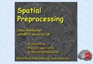
Reorientation and registration demo • Now to SPM… • … for more detail on things like mutual information, please see the May 2011 slides and/or my video athttp://www.fil.ion.ucl.ac.uk/spm/course/video/
B-spline Interpolation A continuous function is represented by a linear combination of basis functions 2D B-spline basis functions of degrees 0, 1, 2 and 3 B-splines are piecewise polynomials Nearest neighbour and trilinear interpolation arethe same as B-spline interpolation with degreesof 0 and 1.
Motion in fMRI • Is important! • Increases residual variance and reduces sensitivity • Data may get completely lost with sudden movements • Movements may be correlated with the task • Try to minimise movement (don’t scan for too long!) • Motion correction using realignment • Each volume rigidly registered to reference • Least squares objective function • Realigned images must be resliced for analysis • Not necessary if they will be normalised anyway
Residual Errors from aligned fMRI • Slices are not acquired simultaneously • rapid movements not accounted for by rigid body model • Image artefacts may not move according to a rigid body model • image distortion, image dropout, Nyquist ghost • Gaps between slices can cause aliasing artefacts • Re-sampling can introduce interpolation errors • especially tri-linear interpolation • Functions of the estimated motion parameters can be modelled as confounds in subsequent analyses
Spatial Normalisation - Reasons • Inter-subject averaging • Increase sensitivity with more subjects • Fixed-effects analysis • Extrapolate findings to the population as a whole • Mixed-effects analysis • Make results from different studies comparable by aligning them to standard space • e.g. The T&T convention, using the MNI template
Standard spaces The Talairach Atlas The MNI/ICBM AVG152 Template The MNI template follows the convention of T&T, but doesn’t match the particular brainRecommended reading: http://imaging.mrc-cbu.cam.ac.uk/imaging/MniTalairach
Normalisation via unified segmentation • MRI imperfections make normalisation harder • Noise, artefacts, partial volume effect • Intensity inhomogeneity or “bias” field • Differences between sequences • Normalising segmented tissue maps should be more robust and precise than using the original images ... • … Tissue segmentation benefits from spatially-aligned prior tissue probability maps (from other segmentations) • This circularity motivates simultaneous segmentation and normalisation in a unified model
Summary of the unified model • SPM8 implements a generative model • Principled Bayesian probabilistic formulation • Gaussian mixture model segmentation with deformable tissue probability maps (priors) • The inverse of the transformation that aligns the TPMs can be used to normalise the original image • Bias correction is included within the model
Mixture of Gaussians (MOG) • Classification is based on a Mixture of Gaussians model (MOG), which represents the intensity probability density by a number of Gaussian distributions. Frequency Image Intensity
Non-Gaussian Intensity Distributions • Multiple Gaussians per tissue class allow non-Gaussian intensity distributions to be modelled. • E.g. accounting for partial volume effects
Modelling inhomogeneity • A multiplicative bias field is modelled as a linear combination of basis functions. Corrected image Corrupted image Bias Field
Tissue Probability Maps • Tissue probability maps (TPMs) are used as the prior, instead of the proportion of voxels in each class ICBM Tissue Probabilistic Atlases. These tissue probability maps are kindly provided by the International Consortium for Brain Mapping, John C. Mazziotta and Arthur W. Toga.
Deforming the Tissue Probability Maps • Tissue probability images are warped to match the subject • The inverse transform warps to the TPMs
Optimisation • Find the “best” parameters according to an “objective function” (minimised or maximised) • Objective functions can often be related to a probabilistic model (Bayes -> MAP -> ML -> LSQ) Global optimum(most probable) Objective function Local optimum Local optimum Value of parameter
Optimisation of multiple parameters Optimum Contours of a two-dimensional objective function “landscape”
Segmentation results Spatially normalised BrainWebphantoms(T1, T2, PD) Tissue probability maps of GM and WM Cocosco, Kollokian, Kwan & Evans. “BrainWeb: Online Interface to a 3D MRI Simulated Brain Database”. NeuroImage 5(4):S425 (1997)
Spatial normalisation results Affine registration Non-linear registration
Spatial normalisation – Overfitting Without regularisation, the non-linear spatial normalisation can introduce unwanted deformation Affine registration (error = 472.1) Template image Non-linear registration without regularisation (error = 287.3) Non-linear registration using regularisation (error = 302.7)
Spatial normalisation – regularisation • The “best” parameters according to the objective function may not be realistic • In addition to similarity, regularisation terms or constraints are often needed to ensure a reasonable solution is found • Also helps avoid poor local optima • Can be considered as priors in a Bayesian framework, e.g. converting ML to MAP: • log(posterior) = log(likelihood) + log(prior) + c
Spatial normalisation – Limitations • Seek to match functionally homologous regions, but... • No exact match between structure and function • Different cortices can have different folding patterns • Challenging high-dimensional optimisation • Many local optima • Compromise • Correct relatively large-scale variability (sizes of structures) • Smooth over finer-scale residual differences
Smoothing • Why would we deliberately blur the data? • Improves spatial overlap by blurring over minor anatomical differences and registration errors • Averaging neighbouring voxels suppresses noise • Increases sensitivity to effects of similar scale to kernel (matched filter theorem) • Makes data more normally distributed (central limit theorem) • Reduces the effective number of multiple comparisons • How is it implemented? • Convolution with a 3D Gaussian kernel, of specified full-width at half-maximum (FWHM) in mm
Example of Gaussian smoothing in one-dimension The Gaussian kernel is separable we can smooth 2D data with two 1D convolutions. Generalisation to 3D is simple and efficient A 2D Gaussian Kernel
References • Friston et al.Spatial registration and normalisation of images.Human Brain Mapping 3:165-189 (1995). • Collignon et al.Automated multi-modality image registration based on information theory. IPMI’95 pp 263-274 (1995). • Ashburner et al.Incorporating prior knowledge into image registration.NeuroImage 6:344-352 (1997). • Ashburner & Friston.Nonlinear spatial normalisation using basis functions.Human Brain Mapping 7:254-266 (1999). • Thévenaz et al.Interpolation revisited.IEEE Trans. Med. Imaging 19:739-758 (2000). • Andersson et al.Modeling geometric deformations in EPI time series.Neuroimage 13:903-919 (2001). • Ashburner & Friston.Unified Segmentation.NeuroImage 26:839-851 (2005). • Ashburner.A Fast Diffeomorphic Image Registration Algorithm. NeuroImage 38:95-113 (2007).
Preprocessing overview Input fMRI time-series Anatomical MRI TPMs Output Segmentation Transformation (seg_sn.mat) Kernel REALIGN COREG SEGMENT NORM WRITE SMOOTH (Headers changed) Mean functional MNI Space Motion corrected ANALYSIS
Preprocessing (fMRI only) Input fMRI time-series TPMs Output Segmentation Transformation (seg_sn.mat) Mean functional Kernel REALIGN SEGMENT NORM WRITE SMOOTH MNI Space Motion corrected ANALYSIS
Preprocessing overview Input fMRI time-series Anatomical MRI TPMs Output Segmentation Transformation (seg_sn.mat) Kernel REALIGN COREG SEGMENT NORM WRITE SMOOTH (Headers changed) Mean functional MNI Space Motion corrected ANALYSIS
Preprocessing with Dartel ... fMRI time-series Anatomical MRI TPMs DARTELCREATE TEMPLATE REALIGN COREG SEGMENT DARTELNORM 2 MNI & SMOOTH (Headers changed) Mean functional Motion corrected ANALYSIS