Fetal Position and Presentaion
460 likes | 643 Vues
Learn to define, diagnose, and manage abnormalities in fetal lie, position, and presentation. Understand the classifications and predisposing factors related to malpresentation. Recognize the types and complications of breech presentation.

Fetal Position and Presentaion
E N D
Presentation Transcript
overview • This lecture discusses how to define, diagnose, and manage the abnormalities of fetal lie, position, and presentation.
Definitions • Fetal attitude: Relationship of fetal head to spine: • flexed, (this is the normal situation) fetuses have a tendency to assume a fully flexed posture • neutral (“military”), • extended.
Definitions • Fetal lie: the relationship between the longitudenal axis of the fetus with respect to the long axis of the mother could be longitudinal lie, transverse lie, oblique lie. • longitudinal, (resulting in either cephalic or breech presentation) • oblique, (unstable, will eventually become either transverse or longitudinal) • transverse (resulting in shoulder presentation).
Definitions • Fetal presentation: to which anatomical part of the fetus is leading, that is, is closest to the pelvic inlet of the birth canal. According to the leading part, this is identified as a cephalic, breech, or shoulder presentation.
Classification of presentation • cephalic presentation (head first): 95% • vertex (crown) — the most common and associated with the fewest complications • sinciput (forehead) • brow (eyebrows) • face • chin
When the head is present in the lower uterine segment “Cephalic” the presentation may be : • Vertex (area between Coronal suture and anterior fontanel) 99% • Face • Brow During the antenatal period It is difficult clinically to diagnose that the presentation is vertex, brow or face so it is used to say cephalic presentation
Vertex 99% Face Brow
Classification of presentation 2. breech presentation (buttocks or feet first): 4% • complete breech • footling breech • frank breech 3. shoulder presentation: 0.5% • arm • shoulder • Trunk 4. Oblique presentation: 0.5%
Definitions • Point of direction: The most dependent portion of the presenting part
Definitions • The fetal position: the location of the point of direction with reference to the pelvis of the mother as viewed by the examiner. position may be right or left as well as anterior or posterior. • Note: fetus enters pelvis in occipito-transverse plane (left or right), descent and flexion then rotates 90 degrees to occipito-anterior (most commonly)
Definitions • Malpresentation: is any presentation other than a vertex presentation (with the top of the head first). In other words: It is the situation where the fetus within the uterus is in any position that is not cephalic "head down".
Predisposing factors to malpresentation • Prematurit • Multiple pregnancy. • Abnormalities of the uterus, eg fibroids. • Partial septate uterus. • Abnormal fetus. • Placenta praevia.
Predisposing factors to malpresentation • Oligohydramnios • Large Fetus • Large Fetal head • Congenital Abnormalities • Cord around the neck
Thr problem in malposition and malpresentation is the fetus is in an abnormal position or presentation that may result in prolonged or obstructed labour.
Breech presentation • Breech pregnancy is a condition of pregnancyin which the fetus is not in the head-down position in the uterus. • Breech presentation is the most common malpresentation, • by about 36 weeks of pregnancy, the baby should have moved into the head-down position • If this has not happened, it is called a breech presentation.
Incidence • Before term between 28 –36 weeks 10-15 % • After 37 completed weeks 3%
Types of breech presentation There are three types of breech presentation: • Complete: both of the baby's knees are bent and his feet and bottom are closest to the birth canal. • Incomplete: one of the baby's knees is bent and his foot and bottom are closest to the birth canal. • Frank: baby's legs are folded flat up against his head and his bottom is closest to the birth canal.
Breech presentation • Breech presentation is much more common in premature labour. • the baby is positioned with the buttocks down and the head up. • The mother may or may not be aware of any symptoms of a breech pregnancy • Complications include difficult vaginal delivery, fetal distress, birth defects and compression of the umbilical cord.
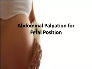
Diagnosis of Breech Presentation • Abdominal examination
Diagnosis of Breech Presentation 2. Vaginal examination when labor is prolonged, the buttocks may become markedly swollen, rendering differentiation of face and breech very difficult; the anus may be mistaken for the mouth.
Diagnosis of Breech Presentation 3. Ultrasound Sonography should ideally be used to confirm a clinically suspected breech presentation Radiation exposure may be reduced considerably by using computed tomographic pelvimetry
Breech presentation • A diagnosis of breech pregnancy and/or breech presentation is made by ultrasound. • In the later stages of pregnancy, it is often possible for a licensed physician or nurse midwife to feel a breech presentation through the wall of a pregnant woman's abdomen.
management • Infants in a breech presentation that are unable to be repositioned into the vertex position are often delivered by cesarean section. • In some cases it is possible to safely deliver an infant vaginally in abreech presentation. • External cephalic version
management Breech allowed to deliver virginally when • No other complication medical or obstetrical with breech • Estimated Fetal size between 2.5 - 3.5 kg • Adequate pelvis
complications • Rupture of fetal membranes • marked molding • cord prolapsed → fetal distress →fetal death • prolonged and complicated labour • Maternal distress → dehydration → keto acidosis • Infection • obstructed labour → uterine rupture →maternal death
complications • Prematurity • Cord prolapse • Asphyxia • intra ventricular hemorrhage • Fetal trauma
indications to the cesarean section • large fetus (the weight of the fetus estimated 3700 g and more). • any degree of contraction or unfavorable shape of the pelvis. • deflexed head. • uterine dysfunction. • previous perinatal death of children suffering from birth trauma. • fetal hypoxia.
External version • attempt to substitute a cephalic presentation by external version. • non-surgical technique to move the baby in the uterus between 37 and 39 weeks • medication ( B-agonists)is given to help relax the uterus. • use of ultrasound to determine the position of the baby • has a high success rate. However
correcting gymnastics • rom 30-32 weeks -correcting gymnastics in breech presentations
Risks of external version • fractured fetal bones • precipitation of labor • premature rupture of membranes • abruptio placentae • fetomaternal hemorrhage (0-5%) • cord entanglement ( <1.5%) • transient slowing of the fetal heart rate
BROW PRESENTATION : • partial extension of the fetal head so that the occiput is higher than the sinciput • On abdominal examination, more than half the fetal head is above the symphysis pubis and the occiput is palpable at a higher level than the sinciput. • On vaginal examination, the anterior fontanelle and the orbits are felt.
Face Presentation • hyper-extension of the fetal head so that neither the occiput nor the sinciput are palpable on vaginal examination • On abdominal examination, a groove may be felt between the occiput and the back. • On vaginal examination, the face is palpated, the examiner’s finger enters the mouth easily and the bony jaws are felt.
Shoulder Presentation • the long axis of the fetus is transverse • The shoulder is typically the presenting part. • On abdominal examination, neither the head nor the buttocks can be. • On vaginal examination, a shoulder may be felt • An arm may prolapse and the elbow, arm or hand may be felt in the vagina.