Download
1 / 33
540 likes | 2.01k Vues
Pancytopenia and Aplastic Anemia. Pancytopenia : Definition. The simultaneous presence of Anemia Leukopenia Thrombocytopenia Hb< 11.5 g/dL (adult females),< 13.5 g/dL (adult males) WBC < 4x10^9/L (4000/mm3) Plt. < 150 x10^9/L (150.000/mm3 ). Aplastic anemia

E N D
Pancytopenia : Definition • The simultaneous presence of • Anemia • Leukopenia • Thrombocytopenia • Hb< 11.5 g/dL (adult females),< 13.5 g/dL (adult males) • WBC < 4x10^9/L (4000/mm3) • Plt. < 150 x10^9/L (150.000/mm3 )
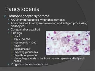
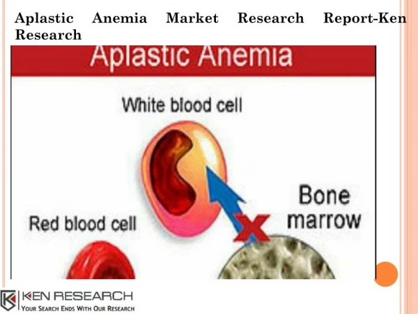
Aplastic anemia Bone marrow infiltration by Hematologic malignancies Non-hematologic Tm met. Storage cell disorders Osteopetrosis Myelofibrosis 3. Paroxysmal nocturnal hemoglobinuria (PNH) 4. Myelodysplastic syndrome 5. Hypersplenism 6. Vit B12 or folate deficiencies 7. S. Lupus erythematosus 8. Cytotoxic agents and antimetabolites 9. Radiotherapy 10. Overwhelming infections 11. other Causes of Pancytopenia
Clinical features • Related to • Pancytopenia Or • Underlying condition/disease
Symptoms/Findings • Related to pancytopenia • The presenting symptoms are related to anemia or thrombocytopenia • Leukopenia may sometimes be life threatining (eg: late in the course of AA, severe neutropenia) • Anemia develops slowly ( long life span of RBC’s) • symptoms of gradual onset.
Symptoms/Findings • Related to pancytopenia • Thrombocytopenic type bleeding may occur and • severity depends on the plt. number. • eg:spontaneous bleeding indicates plt<20.000/mm3 • Infections are related to neutropenia , with severity depending on the decrease in neutrophyl counts.
Symptoms/Findings • Related to the cause of pancytopenia eg: • Splenomegaly: Hypersplenism, lymphoma, leukemia, myelofibrosis etc • Lymphadenomegaly:Lymphoma , leukemia,SLE etc • Atrophic glossitis: Megaloblastic anemia • Others
Investigation of Pancytopenia (Outline) • History: • Age , sex, occupation, diet • Chemical or drug or radiation exposure • Bone pain • Fever, night sweats , malaise, weight loss • Symptoms related to diseases that cause splenomegaly
Investigation of Pancytopenia (Outline) • Physical exam: • Splenomegaly • Bone tenderness • Hepatomegaly • Lymph node enlargement • Gingival hypertrophy • Signs of liver failure or portal hypertension • Evidence of malignancy
Investigation of Pancytopenia (Outline) • Lab. • Essential tests • CBC:Pancytopenia • Reticulocyte count • MCV • Peripheral blood smear • Anizocytosis, poikilocytosis, • leuko-erythroblastosis • neutrophyl granules, • neutrophyl segments, • rouleaux formation, • atypical cells • Bone marrow exam. (aspiration + biopsy)
Investigation of Pancytopenia (Outline) • Lab: • Further investigations when required • X-Rays: Bone, chest etc • Alk. Phosp, acid. Phosp • Serum protein electrophoresis • Anti-DNA, FANA, etc • Urinary proteins (Bence-Jones) • Lymph node or other biopsies
Aplastic Anemia (AA) • The term AA is first used by Ehrlich in 1888 • Describes a disorder of unknown etiology characterized by • pancytopenia with • hypo or acellular bone marrow. • It is one of the stem cell disorders.
Fanconi’s anemia: Autosomal. recessive inheritance Skeletal and renal defects Hyperpigmentation Small stature Hypogonadism Chromosomal changes Familial AA (non-Fanconi) Familial but without features of Fanconi’s anemia Dyskeratosis congenita Skin, nail and hair abnormalities Telangiectasia Alopecia Abnormal sweating Mental retardation Growth failure and hypogonadism Shwachman–Diamond syndrome Classification of AA I-Inherited AA
FA Occurrence of Pancytopenia: Age 7(med) 90% up to age 40 Leukemia & other malignancy hepatic squamous cell carcinomas of the vulva, oesophagus,head and neck Important: Chromosomal breakage and hypersensitivity to the clastogenic effect of DNA cross-linking agents diepoxybutane (DEB) and mitomycin C (MMC)
Abnormal skin pigmentation Nail dystrophy Bone marrow failure > 80-90 % up to age 30 Leucoplakia Learning difficulties Developmental delay mental retardation Pulmonary disease Short stature Extensive dental caries/loss Oesophageal stricture Premature hair loss Malignancy Intrauterine growth retardation Liver disease/peptic ulceration/enteropathy Ataxia/cerebellar hypoplasia Hypogonadism/undescended testes Microcephaly Urethral stricture/phimosis Osteoporosis/aseptic necrosis/scoliosis Deafness Dyskeratosis congenita
DC genetic subtypes • X-linked recessive 40% (Xq2) • Autosomal dominant 5% (3q21–3q28) • Autosomal recessive 50 %
Shwachman–Diamond syndrome • Exocrine pancreatic insufficiency (100%), • Bone marrow dysfunction (100%) and • Other somatic abnormalities (particularly involving the skeletal system) • SDS gene (SBDS) on 7q11 • important role in RNA metabolism and/or ribosome biogenesis.
Shwachman–Diamond syndrome • Short stature (~70%), • Ichthyotic skin rash (~60%). • Metaphyseal dysostosis ~75% • Other abnormalities include • hepatomegaly, • rib/thoracic cage abnormalities, • syndactyly, cleft palate, dental dysplasia, • ptosis and skin pigmentation.
II-Acquired AA Idiopathic Radiation Drugs/chemicals Chloramphenicol NSAID: (phenylbutasone,indomethacin,gold etc) Oral hypoglycemic drugs (chlorpropamide,tolbutamide) Antithyroid drugs, phenothiazines, antimalarials, diuretics,antiepileptics Antineoplastic and cytotoxic drugs Pesticides Solvents and glues: benzene,toluene,xylene,naphtalene Dyes and industrial toxins Others Classification of AA
4. Infections Hepatitis E.Barr virus Rubella CMV HIV Parvovirus Brucellosis Tbc Toxoplasmosis 5. Paroxysmal nocturnal hemoglobinuria(PNH) 6. Immunologic disorders SLE, eosinophilic fasciitis Graft- versus- Host Disease Hypoimmunoglobulinemia Thymoma 7.Pregnancy 8. Pancreatic insufficiency Classification of AA (continued)
Epidemiology of AA • A disease of the young • Median age: about 25 y • 1.5 – 2 /1.000.000-year • Equal sex ratio
Pathophysiology • Defective stroma • Stem cell damage • Damage to stem cell DNA: Radiation, drugs etc • Later progenitor cell damage: Viruses • Immune mediated • Inadequate production of growth factors?
Immune mediated injury • Blood and bone marrow of AA patients supress normal progenitor cell cultures • Cultured AA marrow recovers colony formation after removal of T-cells • Immunosupressive therapy is effective in about half of AA patients • Viruses may cause an infection of the stem cell further leading to an immune response to permanent stem cell failure
Clinical Features of AA • History: • Bleeding • Symptoms of anemia • Infections • Drugs, chemicals or other etiologically important exposures have to be questioned.
Clinical Features of AA • Physical exam: • Petechiae, ecchymosis • Retinal bleeding • Pallor • Fever and other signs of infection • Presence of lymphadenomegaly and /or splenomegaly are unusual (indicate other diagnoses).
Clinical Features of AA • LAB: • Pancytopenia • Reticulocytes: Low or absent • RBC: normochrome-normocytic,or slight macrocytosis • Neutropenia and relative lymphocytosis • Red and white cell precursors are almost never seen in the peripheral smear
Clinical Features of AALAB: • PNH tests may be positive (Ham’s or sucrose lysis tests,others) • Serum iron is increased • Bone marrow : • Aspiration; Dry tap • Biopsy; all three cell lines are reduced or absent, raplaced by fatty tissue, residual lymphocytes, increased iron stores,rarely hot spots of hematopoesis
Course and prognosis of AA • Definition of severe aplastic anaemia: 1-hypocellular bone marrow 2-neutrophils <500/mm3 3-platelets<20.000/mm3 4-reticulocytes<20.000/mm3(<%1) • Survival in severe disease is about 1 year if it is treated with transfusions only. • Very severe AA: Severe AA criteria + Neutrophils:< 200/mm3
Treatment of AA(1) Treatment alternatives: • Allogeneic bone marrow/stem cell transplantation • Immunosupressive treatment • Androgens • Hematopoietic growth factors • Supportive therapy
Treatment of AA (2) • Stem cell transplantation: • Young patients with severe AA and a HLA matched donor should undergo a stem cell transplantation (minimally transfused patients have a chance of > 80% survival ) • Risks: GVHD, engraftment failure, other
Treatment of AA (3) • Immunosupressive treatment • ATG or ALG (antithymosit or antilymphocytic globulin ) • Cyclosporin A • Prednisolone (50% chance of recovery ) • Risks: • Early:infections • Late: MDS, PNH, leukemia, other malignancies, relapse
Treatment of AA (4) Supportive treatment • Hemopoetic Growth Factors: GM-CSF, G-CSF: efficiency ? (limited or not) • Transfusions: • Only when indicated • Exclude family members as transfusion donors • Cellular blood products must be irradiated before transfusions • May cause:Iron overload (Repeated RBC’s), alloimmunization, infection transmission (eg : HIV,HCV,HBV,CMVetc), • Infections:Preventive care, early diagnosis and treatment
Pure Red Cell Aplasia • Anemia + Erythropoetic hypoplasia/aplasia occuring in a normocellular bone marrow Classification • Congenital (Diamond-Blackfan Syndrome) • Acquired • During chronic hemolytic anemia (aplastic crisis) • Infections(eg: Parvovirus B19) • Malnutrition • Drugs (Alpha Methyl Dopa, Azathioprine, Carbamazepine,Gold, NSAID,RMP ,Chloramphenicol etc) • Thymoma • Malignancy • Idiopathic