Comprehensive Overview of Kidney Development | Dr. Shivaram Bhat P.
480 likes | 958 Vues
Explore the intricate stages of kidney development, from embryology to applied aspects, with a focus on the urinary system and nephron formation. An in-depth guide to kidney embryology and anomalies to enhance understanding.

Comprehensive Overview of Kidney Development | Dr. Shivaram Bhat P.
E N D
Presentation Transcript
DEVELOPMENT OF KIDNEY • Dr. Shivaram Bhat P. • Yenepoya Medical college Created – September, 2013
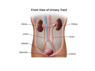
Topic at glance • Urinary system – kidneys, ureters, urinary bladder and urethra. • Embryology – Intermediate mesoderm and Cloaca
OBJECTIVES • Stages of Kidney Development • Development of Collecting System • Development of the Secretory system/ Nephron • Development of Vasculature • Applied Aspects of kidney development
Questions • Embryology of kidney • Anomalies of kidney • Intermediate mesoderm • Nephrogenic cord • Enumerate the anomalies of kidney • Ureteric bud • Metanephros • Ascent of kidneys • Polycystic kidneys
Timeline of Kidney Embryology • Week 4 :Pro nephros, appearance of Mesonephric duct • Week 5 :Meta nephros • Day 28 : formation of Ureteric Bud (UB) • Week 4-8 : Initial MM induction and UB branching • Week 8 : First nephrons are formed • Week 6-8 : kidneys ascend from pelvis to lumbar location • Week 8-15 : Period of UB branching with stochastic formation of UB ampulla and nephron units • Week 10 : kidney function begins • Week 32-36: End of Nephrogenesis
Basic Concepts The 3 embryonic germ layers INTERMEDIATE MESODERM
Mammalian Kidney Development • After the folding of the embryonic disc, the intermediate mesoderm forms a bulging on the posterior abdominal wall, called the NEPHROGENIC CORD/ UROGENITAL RIDGE • It extends from the cervical region to the sacral region of the embryo. UROGENITAL RIDGE
Stages of Kidney Development • The Human Kidney develops in 3 successive stages (rostral to caudal) • PRONEPHROS • MESONEPHROS • METANEPHROS • They are aligned adjacent to the Wolfian / Nephric Duct
Pronephros • The PRONEPHROS develops from the cranial most part of urogenital ridge. • It is transitory and regresses completely by 5 weeks of gestation • Forms the kidney in larval stages of amphibians and fish • It is non functional in Humans.
Mesonephros • MESONEPHROS develops caudal to the Pronephros. • It consists of a series of tubules that drain into the nephric duct, which can be called the Mesonephric duct. • Excretory organ for embryo until metanephros takes over. • By the 4th month of gestation-completely disappears. • Before its degeneration some of its cells migrate and ultimately form the • Adrenal glands • Gonads
Metanephros • METANEPHROS, third and final stage of Kidney development • It results from Reciprocal inductive signals between the Metanephric Mesenchyme (MM) and the Ureteric Bud (UB) at the caudal end of the Urogenital bridge. • Ureteric bud is an outgrowth at the distal end of the Wolfian duct, first visible at approx. 5 weeks of gestation.
Development of the Collecting System • The collecting ducts are all derived from the Ureteric Bud. • The ureteric bud initially penetrates the metanephric mesoderm, and then undergoes repeated branching to form the ureters, renal pelvis, major calyces, minor calyces, and collecting ducts. • This branching is highly patterned
After the first few rounds of branching of the UB derivatives and the concomitant induction of nephrons, the kidney begins to become divided into an outer CORTICAL region (where nephrons are being induced) and an inner MEDULLARY region where the collecting system forms. • As growth continues successive groups of nephrons are induced at the peripheral regions of the kidney, known as the NEPHROGENIC ZONE.
Development of Nephron • The METANEPHRIC MESENCHYME (MM) becomes distinct from the surrounding loose mesenchymal cells and come to lie adjacent to the URETERIC BUD (UB). • Upon invasion of UB in the loose MM, signals from MM cause UB to branch into a T - tubule • Signals from UB induce the MM to condense along the surface of UB
After condensation a subset of the MM aggregates inferior and adjacent to the tips of the branching ureterIc bud, forming the PERITUBULAR AGREGATES. • These undergo mesenchyme – to – epithelial transformation and form the RENAL VESICLE • The Renal vesicle ultimately forms the • Glomerulus • Proximal Convoluted tubule • Loop of Henle • Distal Convoluted tubule Renal Vesicle
Renal Vesicle in contact with the epithelium of the ureteric bud forms the ‘COMMA SHAPE’ • Glomerulus develops from the most proximal end of the Renal Vesicle, farthest from UB tip. • These cells develop into Podocytes. • Vascular cleft develops between podocyte layes and the more proximal cells – ‘S SHAPE’ • Endothelial cells migrate into the vascular cleft • Mesangial cell ingrowth follows the endothelial cells into the vascular cleft.
Development of Vasculature • VASCULOGENESIS – de novo differentiation of previously non vascular cells into structures that resemble capillary beds. • ANGIOGENESIS – sprouting of cells from early capillary beds to form mature vessel structures. • Endothelial progenitors withing the MM give rise to renal vessels in situ, these capillaries form a rich network around the developing nephric tubules.
Renal Ascent The fetal metanephros is located at vertebral level S1-S2, whereas the definitive adult kidney is located at vertebral level T12-L3. From 6th to 9th weeks: kidneys ascend to a lumbar site just below adrenals As the kidneys migrate, they are vascularized by a succession of transient aortic sprouts that arise at progressively higher levels final pair forms in the upper lumbar region and becomes the definitive renal arteries occasionally, a more inferior pair of arteries persists as accessory lower pole arteries Initially the kidneys face anteriorly, but during the ascent, the kidneys rotate 90°causing the hilum to finally face medially.
Speculated that the differential growth of the lumbar and sacral regions of the embryo plays a role
Anomalies in number • Renal agenesis When ureteric bud fails to contact MM • Duplications When ureteric bud splits early before reaching MN • Ectopic ureters When two ureteric buds arise from mesonephricduct
Anomalous Ascent • When the kidney fails to ascend properly, its location becomes ectopic. • The inferior poles of the kidneys may also fuse, forming a horseshoe kidney and ascent is stopped by inferior mesenteric artery • Kidney may fuse to the contralateral one and ascends to the opposite side, resulting in a cross-fused ectopy.
Anomalous position • Malrotated Kidneys - Calyces face anteriorly or antrolaterally. Have some element of obstruction causing inadequate drainage – leading to infection & stone formation Other abnormal positions include- • Ventral Position • Ventromedial Position • Dorsal Position • Lateral Position
Polycystic kidneys • Hereditary – autosomal dominant • Not manifested before 30 • Kidneys enlarged, studded with cysts • Unyeilding capsule compresses renal parenchyma causing atrophy • Liver,lungs and pancreas may be affected • Defact : not clear, many theories
Polycystic kidneys • Loin pain- weight dragging upon peddicle or capsule stretch, hemorhage in cyst, stone • abdominal mass- confused with cystic tumor • hematuria- cyst rupture in pelvis,moderate, episodic. • hypertention, infection, & uremia/CRF. • Nonspecific symptoms: anorexia, headache, vague abdominal discomfort, vomiting, drowsiness, anemia. • ESRD: suddenly in middle age, survival without RRT ( dialysis/ transplant) unlikely