Download
1 / 58
590 likes | 698 Vues
Learn about the sequential stages of tooth development from initiation to crown and root formation, and potential malformations. Explore key processes such as enamel and dentinogenesis, epithelial-mesenchymal interactions, and the differentiation of ameloblasts. Discover how anomalies can affect this intricate process.

E N D
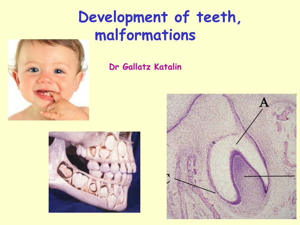
Development of teeth, malformations Dr Gallatz Katalin
DEVELOPMENT OF THE CROWN DEVELOPMENT OF THE ROOT ERUPTION MALFORMATIONS
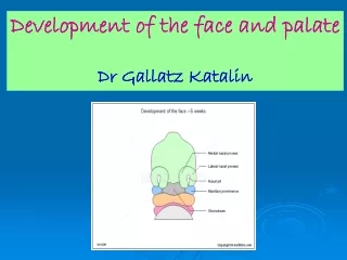
The development of the teeth starts at 6th week with the proliferation of the ectoderm of the stomodeum
The toothdevelopmentstartsinthe 6thweek, withtheformation of the dentallaminafromtheoralepithelium. Dental lamina -Thedentallaminaes(U-shapedbands) follow thecurves of theprimitivejaws.
Dentallamina • Eachdentallaminadevelopstencenters of proliferation, thetoothbuds of thedecidous teeth. • The toothbuds of thepermanentteeth appearlateronthepalatineorlingualaspect of the decidousteeth
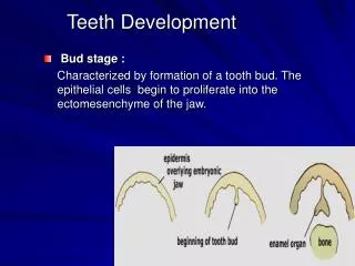
1 TOOTH DEVELOPMENT 2 • initiation stage – 6th to 7th week • bud stage – 8th week • cap stage – 9th to 10th weeks • bell stage – 11th to 12th weeks EO – enamelorgan • apposition stage • maturation stage 3 EO 4
CAP STAGE BELL STAGE ENAMEL ORGAN DENTAL SAC The mesenchyme invaginates into the tooth buds resulting thecap stagethan thebell stage
CAP STAGE • Enamelorgan • Dental papilla • Dentalsac (follicle)
PARTS OF THE DEVELOPING TEETH ECTOMESENCHYME (from the neural crest) ECTODERM Enamel organ Dental papilla Dental sac DENTINCEMENT PULPPERIODONTAL LIG. ALVEOLUS ENAMEL
DS DP
DENTAL SAC ENAMEL ORGAN DENTAL PAPILLA
Parts of the developing tooth enamel organ dental papilla dental sac ameloblasts – enamel producing cells differentiate from the inner enamel epithelium odontoblasts– dentin producing cells differentiate from the ectomesenchymal cells of the dental papilla (neural crest origin) enamel knot
ENAMEL KNOT – SIGNAL CENTER *Non-dividingcellsfromtheinner enamelepithelium, • producedifferentsignalmolecules, whichtransporttheinformation betweentheectodermal and ectomesenchymalcells BMP, FGF,ShH, activin enamel knot
Tooth formation: Initial stages - development of the dental lamina - neural crest cells migrate into the developing mesenchyme - a basement membraneseparates the developing oral epithelium and mesenchyme The initiation of tooth formation starts around the 37th day of gestation.
CAP STAGE • Tooth bud proliferates and forms a cap shaped organ • this stage marks the beginning of histodifferentiation (differentiation of tissues) • the tooth germ also begins to take on form – start of morphodifferentiation • enamel organ is formed – produces the future enamel (ectodermal origin)
CAP STAGE Below this cap is a condensing mass of mesenchyme(ectomesenchyme) – dental papilla – produces the future dentin and pulp tissue. The basement membrane separating the enamelorgan and the dental papilla becomes the future site for the dentinoenamel junction (DEJ) • The mesenchyme surrounding the enamel organ is the dentalsac
CAP STAGE • enamel organ + dental papilla + dental sac is considered the developing tooth germ • tooth germs are found in the developing dental arches - primary dentition • Enamelorgan • Dental papilla • Dentalsac (follicle)
Bell Stage • Continuation of histodifferentiation and morphodifferentiation • differentiation produces 4 celllayer within the ENAMEL ORGAN • 1. inner enamel epithelium • 2. outer enamel epithelium • 3. stellate reticulum • 4. stratum intermedium
DENTAL PAPILLAundergoes differentiation and produces two types of cells 1. outer cells of the DP–ectomesenchymal cellsdifferentiateinto dentin-secreting cells odontoblasts 2. central cells of the DP form the connectivetissue of the pulp Dental sac/follicle increases its collagen content and differentiates at a later stage than the EO and DP Bell Stage
APPOSITION MATURATION DENTINOGENESIS: firsttheodontoblastsproducepredentin (organicmaterial) mineralization AMELOGENESIS: thefirstlayer of predentininduces theinnerenamelepithelialcells, theydifferentiateinto ameloblasts and secretthe organicmatrix of theenamel, mineralization zománc szekréció dentin szekréció ECTO-MESENCHYMAL INTERACTIONS
5 4 6 3 APPOSITIONAL STAGE 1 1. oral ep. 2. outer enamel ep. 3. stellate reticulum 4. inner enamel ep. 5. dental papilla 6. cervical loop 2 blood vessels http://www.iob.uio.no/studier/undervisning/histologi/index.php
5 4 6 7 9 1 8 3 2 APPOSITIONAL STAGE 1. dental papilla 2. preameloblasts 3. Preodontoblasts 4. odontoblasts 5. predentin 6. ameloblasts 7. dentin 8. stratum intermedium 9. enamel http://www.iob.uio.no/studier/undervisning/histologi/index.php
Amelogenesis – Ameloblasts (enamelproducingcells) 1.phase: granularprocessappearsontheapicalsurface of theameloblasts theTomes-process, itcomesoffand formstheorganicmatrix 2. phase: calcification (mineralisation and maturation) 4 micrometerthickenamellayer/day Retziuslines.
Stages of theamelogenesis 1. stage of the secretion 2. stage of the preabsorption 3. stage of the maturation
Summary of the development of the crown 1.Formation of the dental lamina 2.Formation of the enamel organ and the preameloblasts 3.Formation of the dental papilla and odontoblasts 4.Secretion of the predentin 5.Differentiation of the ameloblasts amelogenesis 6.Mineralization of the dentin mineralization of the enamel 7.End of the amelogenesis secretion of the Nasmyth’s membrane
TOOTH DEVELOPMENT Development of the crown
DEVELOPMENT OF THE ROOT HERTWIG’S ROOT SHEATH
HERTWIG-root sheath Composed by the inner and outer enamel epithelium
DEVELOPMENT OF THE ROOT 1. Formation of the Hertwig’sroot sheath 2. Formation of the odontoblasts
DEVELOPMENT OF THE ROOT 3.dentinogenesis 4.desintegration of the Hertwig’s sheath 5.formation of the epithelial islands of Malassez
DEVELOPMENT OF THE ROOT 6. cementogenesis
1 mm ERUPTION • Axial movement toward oral epitheliumstarts when the root develops. http://www.anat.ucl.ac.uk/research/arnett_lab/
Changes in epithelium during eruption enamel cuticle oral ep. junctional ep. reduced enamel ep.
ERUPTION Alveolar bone and connective tissue are resorbed as teeth erupt. osteoclasts http://www.anat.ucl.ac.uk/research/arnett_lab/