IMAGING MODALITIES
IMAGING MODALITIES. Computerized Tomography Magnetic Resonance Imaging. Advantages of C.T. Detection of calcification and calvarial defects No contraindication. Disadvantages of CT. Use of ionizing radiation Reaction to iodinated contrast Nephrotoxicity. Advantages of MRI. No radiation

IMAGING MODALITIES
E N D
Presentation Transcript
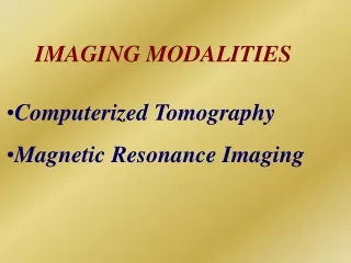
IMAGING MODALITIES • Computerized Tomography • Magnetic Resonance Imaging
Advantages of C.T • Detection of calcification and calvarial defects • No contraindication
Disadvantages of CT • Use of ionizing radiation • Reaction to iodinated contrast • Nephrotoxicity
Advantages of MRI • No radiation • Excellent soft tissue resolution • Multiplanar imaging
Limitations of MRI • Hyperacute bleed • Evaluation of calcification
Contraindications for MRI • Cochlear implants • Cardiac pace maker • Neuro stimulator
Lissencephaly • Most severe of neuronal migrational • abnormalities • Generalized paucity of gyral and sulcal • formation • Vertically oriented sylvian fissures
Torch Infections • Toxoplasmosis • Rubella • Cytomegalovirus - Most common • cause of congenital CNS infection • Herpes simplex virus
Focal cortical dysplasia • Common location – temporal lobes • Expanded gyrus with abnormally oriented • sulci and thickened cortex • Subcortical white matter hyperintensity • Surgical excision of dysplastic focus when • possible is often curative
Cortical dysplasia – Balloon Cell Type of Taylor • Focal cortical thickening • Blurring of the gray-whitematter junction • Hyperintensity (on T2-weighted images)of • subcortical white matter often tapering • toward the ventricle
Unilateral megalencephaly • Hamartomatous overgrowth of a part or whole of cerebral hemisphere • Ipsilateral migrational defects • Hypoplastic / hyperplastic white matter • Intractable seizures, hemiplegia and severe developmental delay
Peri-Sylvian syndrome • Anomalous cortical development overlying • underdeveloped sylvian fissures • Dorsal perirolandic extension of sylvian • fissures
Septo-optic dysplasia (de Morsier syndrome) • Partial or complete absence of septum • pellucidum • Squared off appearance of frontal horns • Hypoplasia of optic nerves and chiasm • ( 40-80%) • Hypoplasia of hypothalamus
Tuberous sclerosis(Bourneville disease) • Incidence -- 1:10,000-50,000 • Inheritance -- autosomal dominant -- low penetrance -- chromosomes: 9, q32 - 34; 11, ??
Tuberous sclerosis Clinical - “classic” triad of: > Papular facial lesions > seizures > mental retardation- 50% of patients
Tuberous sclerosis • CNS lesions -Subependymal nodules - Giant cell astrocytoma - Cortical tubers - White matter lesions • Non- CNS lesions - Skin, kidneys, cardiovascular, Liver, spleen, pancreas and Musculoskeletal
Sturge-weber syndrome (Encephalotrigeminal angiomatosis) • Inheritance : none • Clinical : port wine stain in CN - V distribution
Sturge-weber syndrome • Aetiology - Normal cortical venous drainage fails to develop • Pathology - Leptomeningeal angiomatous vascular plexus with secondary dystrophic cortical changes
Sturge-weber syndrome • Calcification • Atrophy • Enlarged med, sub-epen veins • Ocular lesions
Periventricular leukomalacia • Commonly seen in premature infants • Ischemic lesions are most obvious in parieto-occipital regions • Paucity of white matter in the parieto-occipital regions • Indentation of the lateral ventricles
Hippocampus Mean volumes: Right Left Male - 2.20+0.47cu.cm 2.17+0.72cu.cm Female- 2.27+0.47cu.cm 2.23+0.48cu.cm Hippocampal sclerosis: 1.46+0.60cu.cm
Hippocampus Normal: NAA/Cho:1.20 + 0.27 Hippocampal sclerosis: NAA/Cho:0.99 + 0.14
Hippocampus T2 Relaxometry Mean T2 time:110-115ms Prolonged in Hippocampal sclerosis
Dysembryoplastic Neuroepithelial Tumor • Slow growing superficial lesions usually within temporal lobe but always supratentorial • Focal cortical lesion , hypointense on T1 & • hyperintense on T2 Wt.images • Surgery is curative