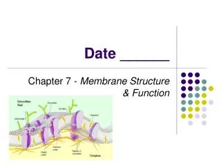
Chapter 10: Membrane Structure
Chapter 10: Membrane Structure. Membrane Structure. Membrane Function Plasma membrane defines cell and maintains differences btwn cytosol and extracellular environment Defines individual organelles Establish ion gradients

Chapter 10: Membrane Structure
E N D
Presentation Transcript
Membrane Structure Membrane Function • Plasma membrane defines cell and maintains differences btwn cytosol and extracellular environment • Defines individual organelles • Establish ion gradients • Contain proteins act as sensors of external signals allow cell to adapt to changing environment
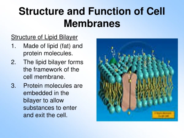
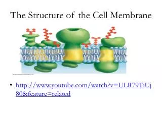
Membrane Structure • General Membrane Structure • Thin film of lipid and protein held together by noncovalent interactions • lipid bilayer serves as basic fluid structure; impermeable barrier • Proteins mediate all other functions of membrane
The Lipid Bilayer Lipids of Membrane • 50% of total membrane mass, 5 x 10⁶/um2 • Amphipathic • Self-sealing • Phosphlipids, cholestrol, glycolipids • Phospholipids= most abundant lipid in membrane • Spontaneously aggregate to bury hydrophobic tail
The Lipid Bilayer Fluidity of Lipid Bilayer • Rapid lateral diffusion of phospholipids 10-8cm/sec • Change places 107 times/sec • Rarely flip-flop • Fluidity depends on composition (phospholipid and cholesterol) and temperature • Phase transition= the temperature at which there is a change of state from liquid to solid
The Lipid Bilayer Fluidity and length and saturation of FA hydrocarbon chains • Short hydrocarbon chain lengths fluidity • Double bonds fluidity
The Lipid Bilayer • Fluidity and cholesterol content Cholesterol fluidity • Provides mechanical stability • 1 cholesterol/phospholipid in eucaryotes • No cholesterol in procaryotes; mechanical stabililty imparted by cell wall
The Lipid Bilayer Major Membrane Phospholipids
The Lipid Bilayer Lipid Rafts • Microdomains enriched in sphingolipids, cholesterol and membrane proteins • Long saturated FA chain of sphingolipids = attractive forces that hold adjacent molecules together • Thicker than other parts of bilayer • able to accommodate membrane proteins concentrating for transport or to enable proteins to function together
The Lipid Bilayer Membrane asymmetry • Phospholipid distribution: phosphatidylcholine and sphingomyelin confined to outer monolayer phosphatidylethanolamine and phosphatidylserine are on inner monolayer • Charge • Proteins • Impt to function (ie, apoptosis and translocation of phosphatidylserine) • glycolipids
The Lipid Bilayer Glycolipids • Sugar containing lipid molecules w/ most extreme assemmetry in distribution • Found exclusively on noncytoplasmic monolayer • On surface of all plasma membranes • gangliosides= most complex, sialic acid containing oligosaccharides, net negative chg, most abundant in pm of nerve cells • Function- protection, cell recognition, transmission of electrical impulses
Membrane Proteins • Proteins Carry Out Specific Functions of Membrane • Give ea type of membrane characteristic functional properties • Amts and types of proteins highly variable • By mass proteins represent 50% lipids 50%; lipids small thus 1 protein/ 50 lipid molecules • Associate w/ membrane in various ways depending on function: transmembrane, integral, peripheral
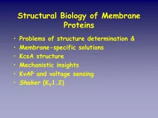
Membrane Proteins Transmembrane Proteins Typically Cross as Alpha Helix • Unique orientation reflection syn and insertion in ER and function • Membrane spanning doamin comprised of nonpolar aa 20-30 • Alpha helix is predominate conformation but beta shees form closed beta barrels that can span membrane as well • Can predict membrane spanning regions via hydropathy plot
Membrane Proteins Beta Barrels form Transmembrane Channels • Tend to be more rigid • Comprised of 8-22 strands • Some pore forming proteins generating water-filled channels for select hydrophilic solutes • Polar side chains line channel on inside; nonpolar side chains project out interact w/hydrophobic core of lipid bilayer
Membrane Proteins • Many Membrane Proteins are Glycolslated • Majority of transmembrane proteins in animal cells are glycolsylated • Sugars added in lumen of ER and golgi • Oligosaccharide chains always present on noncytosolic side
Membrane Proteins • Membrane Proteins can be Solubilized and Purified in Detergents • Distrupt hydrophobic interactions • Bind to hydrophobic regions of membrane and displace lipid molecules • SDS, triton
Membrane Proteins • Red Blood Cells • Model system for studying membranes • Available in lg numbers • Easy to isolate uncontaminated from other cell types • No nucleus or internal organelles • Can prepare ghosts and in-side-out vesicles
Membrane Proteins Ghosts and InSide-Out RBCs Ghosts= empty RBCs prepared by placing cells in soln of low salt to cause water to move into and lyse cells can reseal or be studied while leaky Inside-Out Vesicles
Membrane Proteins Use of Sealed and Unsealed RBC ghosts • Demonstrated some proteins extend across lipid bilayer • Enabled sidedness of proteins to be determined • Label intact sealed ghosts and inside-out vesicles w/ water solutble label that cannot penetrate lipid bilayer– perform SDS PAGE • Exposed internal or external surface to proteolytic enzymes that are membrane impremeant (transmembrane protein will be partially digested from both sides • Labeled antibodies
Membrane Proteins Studies of RBC Plasma Membrane Proteins by SDS PAGE • 15 major protein bands btwn 15,000-250,000 daltons • 3 most prominent comprose more than 65% protein mass; ea arranged differently in membrane • Spectrin • Glycophorin • Band 3
Membrane Proteins Spectrin= Cytoskeletal Protein Assoc. w/ Cytosolic Side of Membrane • Most proteins are peripheral and assoc w/ cytosolic side • Spectrin most abundant • Long, thin, flexible rod 100 nm length, constitutes 25% protein mass • 2.5 x 10⁵ copies/cell • Principle component of cytoskeleton underlying RBC, maintains structural integrity and biconcave shape • Heterodimer formed from 2 lg structurally similar subunits that assoc head to head to form 200 nm long tetramer • Tail ends of 4-5 tetramers linked by binidng short actin filaments and other cytoskeletal components forming meshwork • Abnormalities in spectrin results in anemia and spherical shaped RBCs that are fragile • Ankyrin binds spectrin and band 3 to membrane
Membrane Proteins Spectrin
Membrane Proteins Glycophorin • sm singlepass glycoprotein of 131 aa • Most of mass on external surface of membrane • 100 sugar residues on 16 diff side chains which account for 60% protein mass • >90% sialic acid most of negative chg on surface of RBC • 1 million molec/cell • Function unknown
Membrane Proteins Band 3 • Multipass membrane protein • Catalyzes couple transport of anions • 930 aa thought to extend across bilayer 12X • Allows HCO3 to cross membrane in exchg for Cl- increasing amt of CO2 delivered to lungs
Membrane Proteins • Bacteriorhodopsin is a Proton Pump • Halobacterium salinarum “purple membrane” • 7 transmembrane helices of 25 aa • Retinal light absorbing chromophore linked to lys side chain • Light excites chromophore causing conf chg which results in transfer of H+ from inside to outside cell • Electrochemical gradient used to syn ATP • Can pump up to 100 H+/sec
Membrane Proteins Membrane Proteins Often Function as Lg Complexes
Membrane Proteins Membrane Proteins Diffuse in Plane of Membrane but Do Not Flip-Flop
Membrane Proteins Techniques for Measuring the Lateral Diffusion of Membrane Proteins
Membrane Proteins Confining Proteins and Lipids to Specific Regions of the Membrane
Membrane Proteins • Cell surface Coated with Sugar Residues • Glycocalyx= CHO rich region on cell surface • Proteoglycans= long polysaccharide chains linked to protein core • Oligosahharides also linked to proteoglycans • Possbile functions: protection, receptors, electrical conductivity