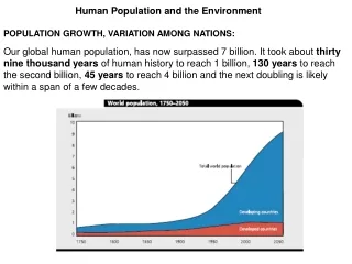
Download
1 / 24
240 likes | 408 Vues
Acta Biophysica Romana 2008. A XAS Study of the Sulphur Environment Location in Human Neuromelanin and Synthetic Analogues. P.R. Crippa, M. Eisner, S. Morante, F. Stellato , F. Vicentin, L. Zecca. Outline. Parkinson’s Disease Neuromelanin X-Ray Absorption Spectroscopy Experiments

E N D
Acta Biophysica Romana 2008 A XAS Study of the Sulphur Environment Location in Human Neuromelanin and Synthetic Analogues P.R. Crippa, M. Eisner, S. Morante, F. Stellato, F. Vicentin, L. Zecca
Outline • Parkinson’s Disease • Neuromelanin • X-Ray Absorption Spectroscopy Experiments • Conclusions
Parkinson’s Disease • Parkinsons’s Disease (PD): • Progressive and fatal neurodegenerative disease • Described in 1817 by James Parkinson • Affects 1-2% of over 50 population • <10% of PD is familial majority of cases are sporadic • 5 clearly defined genetic causes B. Thomas et al.(2007) Hum Mol Gen 16,R183.
PD Pathogenesis Dopamine and acetylcholine are neurotrasmitters that control body movement synapse nerve terminal synaptic vesicle dopamine Dopamine is produced in a small area in the base of the brain, called substantia nigra In Parkinson’s disease NM pigmented neurons die
Neuromelanin Neurons containing neuromelanin pigment • Neuromelanin (NM): • Dark pigment present in neurons of different brain areas • Mixture of similar polymers which are made up of different structural units • Accumulates with aging • Contributes to the protection of neurons from oxidative processes • Pigmented neurons are lost in Parkinson’s disease • Relation between neuronal vulnerability and presence of NM still unclear
NM structure Polymeric compound composed by indolebenzothiazine groups XRD: multilayer (graphite-like) three dimensional structure with planar overlapped sheets consisting of cyclic molecules of indolebenzothiazine ring 15% Covalent bound peptide component 20% lipidic component NM Contains S Binds Fe and Zn L. Zecca et al.(2000) J Neurochem 74, 1758.
Sulphur Content Indolebenzothiazine Cysteine Indolebenzothiazine groups contain S S The peptidic part contains Cysteine (about 3% in weight) No Methionine detected
XAS Experiments X-ray Absorption Spectroscopy (XAS) study at the S K-edge Measurement of X-ray Absorption coefficient m(E) • XAS features • Selective for the absorber • Local probe (~5 Å) • No crystallization needed
Experimental Setup X-ray source IF measurement Solid State Detector Monochromatic beam White beam X-ray mirror Sample Monochromator Ionization chambers Synchrotron I0 and I measurement Incident energy selection
XAS spectrum c(k) X-ray Absorption Near Edge Spectroscopy XANESregion EXAFSregion Extended X-ray Absorption Fine Structure m(E) k E XANESEXAFS EXAFS spectra are analyzed in terms of EXAFS region can be analyzed in the single scattering approximation: characteristic of atomic type indistinguishable for light atoms (N, O, C) introduce multiple scattering terms .
Samples Cerebellum • 6 powder samples • Human Neuromelanin (HNM) • extracted from cerebellum • 3 Synthetic Melanins prepared with different procedures • 2 Model compounds (Cysteine and Trichochrome)
Model Compounds Trichochrome Cysteine Trichochrome S is present as heteroatom in aromatic rings Cysteine S is present in the amino acid side chain
Synthetic Melanins Model of Synthetic Melanin Synthetic compounds similar to natural melanins Enzymatic Oxidation (With Tyrosinase) Dopamine + Cysteine DEC Dopa + Cysteine Pheomelanin Auto-oxidation Dopamine + Cysteine DAC Dopamine Dopa Cysteine
Data Collection Carbon foil Monochromatic beam Sample White beam Monochromator Electrometer Synchrotron • Spectra collected at the D04B • bending magnet beam line of the • Brazilian Synchrotron Light Laboratory • Total Electron Yield (TEY) I0: incident current measured with a 0.75 μmcarbon foil I: sample current collected with an electrometer m=I/I0
Total Electron Yield Auger effect (Non-radiative de-excitation) -The photo-electron is emitted -The core hole is filled by an electron of an upper level -The energy is used up to eject an Auger electron -TEY: detection of all electrons emitted by the sample Fluorescence yield is low for low-Z elements (for S F<0.1) Xs, XA: emission probabilities of fluorescence photon and Auger electron
Results HNM & Model Compounds Model Compounds Cysteine – Trichochrome Significantly different spectral features Natural Melanin HNM different from both
HNM & Synthetic Melanins Synthetic Melanins DAC DEC -Pheomelanin Similar spectral features Natural Melanin HNM Similar to Pheomelanin and DEC
Difference Spectra Qualitative findings are consistent with quantitative analysis of difference spectra
Data Analysis 1- Determination of edge energy E0 2- Spectra shifted by E0 3- Identification of two peaks (white line and first peak) in all spectra 4- P1, P2: positions of the two peaks 5- A1, A2: amplitudes of the two peaks P1 P2 E0
Data Analysis E0 and P1 are the same in all spectra P2 is the same in all spectra but Cysteine
Fit Fits of HNM are obtained minimizing
—— HNM —— Fit HNM = 64% Cysteine + 36% Trichochrome
—— Pheomelanin —— Fit Pheomelanin = 55% Cysteine + 45% Trichochrome
Conclusions We have performed a structural study on natural Neuromelanin and Synthetic Analogues • Identification of percentage of Trichochrome-like and a Cysteine-like components in Human Neuromelanin • S structure is similar in Human Neuromelanin and Synthetic Melanins Pheomelanin and DEC