
3D Structures of LRR-Containing Proteins: Insights into Brassinosteroid Recognition and Inhibition
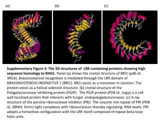
This figure illustrates the three-dimensional structures of leucine-rich repeat (LRR) proteins with high sequence homology to RHG1. Panel (a) presents the crystal structure of BRI1 (PDB ID: 3RGX), crucial for brassinosteroid recognition via its LRR domain and existing as a monomer with a helical solenoid structure. Panel (b) depicts the PGIP protein (PDB ID: 1OGQ), which functions in plant defense by interacting with fungal enzymes. Panel (c) showcases the structure of the porcine ribonuclease inhibitor (PRI, PDB ID: 2BNH), demonstrating its horseshoe configuration and role in regulating RNA levels.

3D Structures of LRR-Containing Proteins: Insights into Brassinosteroid Recognition and Inhibition
E N D
Presentation Transcript
(a) (b) (c) Supplementary Figure 3: The 3D structures of LRR containing proteins showing high sequence homology to RHG1. Panel (a) shows the crystal structure of BRI1 (pdb id: 3RGX). Brassinosteroid recognition is mediated through the LRR domain of BRASSINOSTEROID-INSENSITIVE 1 (BRI1). BR1I exists as a monomer in solution. The protein exists as a helical solenoid structure. (b) crystal structure of the Polygalacturonase inhibiting protein (PGIP). The PGIP protein (PDB id: 1ogq) is a cell wall localized protein that interacts with fungal endopolygalacturonases. (c) X-ray structure of the porcine ribonuclease inhibitor (PRI). The Leucine rich repeat of PRI (PDB id: 2BNH) forms tight complexes with ribonucleases thereby regulating RNA levels. PRI adopts a horseshoe configuration with the LRR motif composed of repeat beta-loop helix units.