Download
1 / 82
830 likes | 1.39k Vues
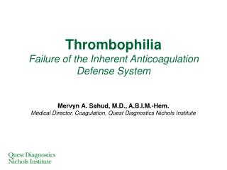
Thrombophilia Failure of the Inherent Anticoagulation Defense System. Mervyn A. Sahud, M.D., A.B.I.M.-Hem. Medical Director, Coagulation, Quest Diagnostics Nichols Institute. Outline. Thrombophilia ( impact on DVT and PE ) Risk factors Coagulation cascade Thrombophilia testing Antithrombin

E N D
ThrombophiliaFailure of the Inherent AnticoagulationDefense System Mervyn A. Sahud, M.D., A.B.I.M.-Hem. Medical Director, Coagulation, Quest Diagnostics Nichols Institute
Outline • Thrombophilia (impact on DVT and PE) • Risk factors • Coagulation cascade • Thrombophilia testing • Antithrombin • Protein C • Protein S • Activated protein C resistance (APCR) • Prothrombin (factor II) mutation • Lupus anticoagulant • Factor VIII excess • Case studies • Recommended work-ups
Outline • Thrombophilia (impact on DVT and PE) • Risk factors • Coagulation cascade • Thrombophilia testing • Antithrombin • Protein C • Protein S • Activated protein C resistance (APCR) • Prothrombin (factor II) mutation • Lupus anticoagulant • Factor VIII excess • Case studies • Recommended work-ups
Valve Cusp Thrombus (Autopsy Specimen)
2 million Americans affected with DVT each year • 600,000 of these develop pulmonary embolism (PE) • 60,000 of those with DVT and PE die each year DVT & PE DVT only Die from PE Thrombophilia DVT, deep vein thrombosis. Rosendall FR. Lancet. 1999:353:1167-1173. Anderson FA Jr, et al. Arch Intern Med. 1991;151:933-938.
Thrombophilia • 5% - 8% of population affected by genetic defects leading to thrombosis predisposition • 25% suffer chronic swelling, skin ulceration, and impaired mobility secondary to “venous hypertension”
DVT…What’s New? • 50% (soon to be 100%) of “unprovoked” DVT cases associated with hereditary thrombophilia • 60% of DVT cases in pregnant women associated with factor V Leiden mutation (A1698G) • DVT often associated with multiple genetic and acquired risk factors • DVT responds to prolonged anticoagulant therapy (eg, low molecular weight heparin) Bucciarelli P, et al. Arterioscler Thromb Vasc Biol. 1999;19:1026-1033.
DVT…What’s New? • New anticoagulants available for non-responsive patients and those who experience side-effects from standard anticoagulants • Family members request DVT screening prior to being placed in a “high-risk” situation • Elevated D-dimer level often indicates thrombosis • Normal D-dimer level has strong NPV NPV, negative predictive value..
What Else is New? • Pulmonary embolism • 4 million patients present to U.S. emergency departments with shortness of breath each year • Shortness of breath = heart failure or pulmonary embolism (PE) • 60% of patients who die in the hospital have PE • PE diagnosis missed in 70% of hospital cases • 10% of patients with acute PE die within first 60 minutes Clagett GP. Chest. 1998;114(Suppl 5):531S-560S.
Thrombophilia Is Often MultigenicMultiple risk factors raises the risk of thrombosis
Outline • Thrombophilia (Impact on DVT and PE) • Risk factors • Coagulation cascade • Thrombophilia testing • Antithrombin • Protein C • Protein S • Activated protein C resistance (APCR) • Prothrombin (factor II) mutation • Lupus anticoagulant • Factor VIII excess • Case studies • Recommended work-ups
Clinical DVT Risk Factors • Surgery for malignant disease • Postoperative sepsis • Major medical illness • Heart failure • Inflammatory bowel disease • Sepsis • Myocardial infarction • Stroke • Age >60 y • Extensive surgery* • Marked immobility, pre- or postoperative • Major orthopedic surgery (eg, hip, knee) • Fracture of pelvis, femur, or tibia *Risk of postoperative thrombosis increases as patient’s age and surgery duration increases; risk also increases with presence of varicose veins and obesity.
Other (<1%) LA (3%) Antithrombin (4%) Unknown (5%) Factor VIII excess (30%) Protein S (10%) Protein C (10%) Hyperhomocysteinemia (10%) APCR, Factor V mutation (20%) Prothrombin (factor II) mutation (15%) Frequency of Biological Risk Factors
Synergistic Effects of Thrombotic Risk Factors aProthrombin 20210GA mutation.
Synergistic Effects of Thrombotic Risk Factors aProthrombin 20210GA mutation.
Risk of Recurrent Venous Thromboembolism Risk of recurrent venous thromboembolism (VTE) in individuals with a thrombophilic defect relative to risk in those with a first episode of VTE and no thrombophilic defect. Weitz J, et al. Hematology Am Soc Hematol Edu Program. 2004;424-438.
Age of Thrombosis Onset in Patients with Common Thrombotic Risk Factors APCR, activated protein C resistance. *May be responsible for venous thrombosis in individuals older than 55-60 years.
Relative Frequency of Various Thrombosis Types Salomon O, et al. Arterioscler Thromb Vasc Biol. 1999;19:511-518.
Outline • Thrombophilia (impact on DVT and PE) • Risk factors • Coagulation cascade • Thrombophilia testing • Antithrombin • Protein C • Protein S • Activated protein C resistance (APCR) • Prothrombin (factor II) mutation • Lupus anticoagulant • Factor VIII excess • Case studies • Recommended work-ups
Coagulation Cascade Crowther MA, Kelton JG. Ann Intern Med. 2003;138:128-134.
Coagulation Cascade Factor VIIa binds to tissue factor (TF) at sites of vascular injury, causing a cascade that ultimately leads to factor IIa (thrombin) generation. Thrombin activates platelets; converts fibrinogen to fibrin; leads to generation of additional thrombin through an autocatalytic loop; converts factor XIII to factor XIIIa, which stabilizes the thrombus; inhibits fibrinolysis; and acts as an anticoagulant by activating protein C. The principal inhibitors of coagulation are activated protein C (which, in concert with protein S, inactivates factors Va and VIIIa), antithrombin (which forms an inactive covalent complex with thrombin and factors Xa, IXa, and XIa), and tissue factor pathway inhibitor (which bonds with and inactivates the tissue factor-factor VIIa complex). Crowther MA, Kelton JG. Ann Intern Med. 2003;138:128-134.
Outline • Thrombophilia (impact on DVT and PE) • Risk factors • Coagulation cascade • Thrombophilia testing • Antithrombin • Protein C • Protein S • Activated protein C resistance (APCR) • Prothrombin (factor II) mutation • Lupus anticoagulant • Factor VIII excess • Case studies • Recommended work-ups
Whom to test? How to test? Why test? Thrombophilia Testing When to test? What to test?
Antithrombin Deficiency Genetic deficiency • Rare • <1% of general population • ~1% - 8% of those with VTE • Heterozygotes • VTE by ~30 years of age • 5- to 50-fold increased risk of VTE • High risk of VTE during pregnancy and postpartum
Antithrombin Deficiency Acquired deficiency • Causes • Liver disease • Malnutrition • Inflammatory bowel disease • Prematurity • DIC • Transfusion reactions • Chemotherapy • Heparin therapy
IIa AT IIa IXa XIIa Xa XIa AT AT AT AT AT AT IIa Role of antithrombin • Direct inhibitor of IIa • Inhibits other factors leading to indirect inhibition of IIa formation Heparin Inactive Complexes
Normal antithrombin III Antithrombin III deficient Therapeutic Monitoring with Antithrombin III
Factors Affecting Test Results OAC, oral anticoagulant.
Protein C Deficiency – Genetic Mechanism Autosomal inheritance • Complete penetrance (dominant mutation) • Heterozygotes • Primary symptom: venous thrombosis • Mild or no penetrance (recessive mutation) • Homozygotes: symptomatic (purpura fulminans) • Heterozygotes: asymptomatic
Protein C Deficiency • Genetic deficiency • Rare • Heterozygotes • <1% of population • ~1-11% of patients with VTE • 30% will have an event during lifetime • ~50% experience VTE by age 40 • Acquired deficiencies • Warfarin, vitamin K deficiency, liver disease, DIC, renal insufficiency
Chromogenic Pro Fewer interfering substances Con Measures cleavage of synthetic substrate Rare mutations not detected by this method Clotting Pro Measures cleavage of natural substrates (factor Va or VIIIa) Con Many interfering substances High factor VIII levels Lupus anticoagulants Heparin >1.0 IU Direct thrombin inhibitors Which Protein C Functional Assay?
Functional Protein C Assay 2 patients missed by clot assay 8 patients missed by chromogenic assay
Protein C Testing Algorithm Protein C Clot Assay Decreased (<70%) Borderline (70% - 75%) Normal or Protein C Antigen Repeat Clot Assay No further testing Decreased (<70%) Increased (70%) Borderline (70% - 75%) Chromogenic Assay Normal adult reference range: 70%
Therapeutic Monitoring with Protein C Normal protein C Protein C deficient
Protein S Deficiency Genetic deficiency • Homozygous (rare) • Purpura fulminans • Heterozygous • < 1% of general population • ~1% to 3% of VTE population • ~50% experience first VTE by 45 years of age Acquired deficiency • Pregnancy and oral contraceptive/HRT use • Inflammation and acute thrombosis
PS Free PS C4b-BP TM IIa IIa TM APC fPS fPS Protein C PC Va or VIIIa TM orVai VIIIai Inactive Cofactors Role of protein C & S • Inhibition of IIa formation
Normal protein S Protein S deficient Therapeutic Monitoring with Protein S
APC Resistance in Patient with Thrombosis APC, when added to plasma from a middle-aged man with recurrent thrombosis, did not result in a prolongation of the clotting time (APC resistance). In contrast, when added to normal plasma (control), APC prolongs the clotting time in a dose-dependent manner. Dahlback B. J Thromb Haemost. 2003;1:3-9.
APC Resistance – Genetic Mechanism • Molecular cause Mutation of APC cleavage site within Factor Va • Mutation site Amino acid #506 (arginine) mutates to a glutamine Amino acid: Arg506→ Gin Nucleotide: CGA→ CAA
APC Resistance – Genetic Mechanism • Molecular function • APC cleaves bond between arginine and glycine • If Arg506 residue is mutated • Bond not cleaved • Factor Va not inactivated by APC • Continued generation of thrombin and clot formation • Thrombosis
Acquired APC Resistance Associated with: • Increased plasma levels of FVIIIa • Presence of antiphospholipid antibodies • 3rd generation oral contraceptives, relative to 2nd generation oral contraceptives • Pregnancy
Prothrombin (Factor II) 20210GA Mutation A, adenine; G, guanine. Zoller B, Svensson PJ, He X, et al. J Clin Invest. 1994;94:2521-2524.
Lupus Anticoagulant Screening • Screening tests • LA sensitive aPTT reagent • Kaolin clotting time (KCT) • Dilute russell viper venom time (dRVVT) • Dilute prothrombin time (dPT) • Minimum of 2 screening tests
Factor VIII Excess O’Donnell J, et al. Thromb Haemost. 1997;77:825-828.
Factor VIII Excess vs Other Thrombophilia Markers aHistory of thromobophilia. O’Donnell J, et al. Thromb Haemost. 1997;77:825-828.
Outline • Thrombophilia (impact on DVT and PE) • Risk factors • Coagulation cascade • Thrombophilia testing • Antithrombin • Protein C • Protein S • Activated protein C resistance (APCR) • Prothrombin (factor II) mutation • Lupus anticoagulant • Factor VIII excess • Case studies • Recommended work-ups