Download
1 / 33
330 likes | 623 Vues
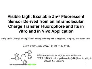
Visible Light Excitable Zn 2+ Fluorescent Sensor Derived from an Intramolecular Charge Transfer Fluorophore and Its in Vitro and in Vivo Application. Fang Qian, Changli Zhang, Yumin Zhang, Weijiang He, Xiang Gao, Ping Hu, and Zijian Guo. J. Am. Chem. Soc. , 2009 , 131 (4), 1460-1468.

E N D
Visible Light Excitable Zn2+ Fluorescent Sensor Derived from an Intramolecular Charge Transfer Fluorophore and Its in Vitro and in Vivo Application Fang Qian, Changli Zhang, Yumin Zhang, Weijiang He, Xiang Gao, Ping Hu, and Zijian Guo J. Am. Chem. Soc., 2009, 131 (4), 1460-1468. NBD:4-amino-7-nitro-2,1,3-benzoxadiazole TPEA:N,N,N’-tris(2 -pyridylmethyl)–N’-(2-aminoethyl)- ethane-1,2–diamine
Zn2+ Plays Vital Roles in Vivo • cellular metabolism • 2. gene expression • 3. Apoptosis • 4. neurotransmission
Schematic Illustration of Putative Zn2+- signaling Pathways • bind to receptors of ion channels of • postsynaptic neurons modulating their activity. • (2) enter postsynaptic neurons via Zn2+- • permeable ion channels. • (3) be taken back up into the presynaptic • neuron and vesicles • (4) diffuse away into the extracellular fluid. Proc. Natl. Acad. Sci. U.S.A. 2003, 100, 3605–3610.
a b ZnAF-2M ZnAF-2 c d ZnAF-3 ZnAF-4 Fluorescence Response of ZnAFs Detecting Extracellularly Released Zn2+ in Hippocampal Slices J. Am. Chem. Soc. 2005, 127, 10197.
Intact in Vivo Imaging of Ca2+ in Zebrafish Larva Maker:Calcium Green-1 AM Pfluegers Arch. Eur. J. Physiol. 2003, 446, 766–773.
What Kinds of Fluorescent Imaging are Essentially Demanded 1.They have no UV-induced phototoxicity and autofluorescence 2.They have no sensor-induced interference visible light excited sensor of biocompatibility is appealing
The Reported Visible Light Excited Zn2+ Fluorescent Sensors fluorescence images of HeLa cells fluorescence images of fibroblast cells (ex)= 400–440 nm (ex)= 356 nm fibroblast cells:皮膚纖維母細胞 HeLa:子宮頸癌細胞 J. Am. Chem. Soc.2004, 126, 712 –713
Novel Approach in Developing Visible Light Excitable Zn2+ Fluorescent Sensor SN2 1.visible ICT 2.lower interference R1=NO2, R2=amine (ex)=460~490 nm 2,1,3-benzoxadiazole (BD) J. Chem. Soc., Perkin Trans. 2 1998, 2165–2173. Biochem. J. 1968, 108, 155–156.
Fluorescence Micrographs of Cells Treated with C6-NBD-X (X=PA, PC, PE)
Chemical Structures of NBD-TPEA, NBD-PMA, and NBD-BPA ICT absorption and large Stokes shift
Intramolecular Charge Transfer (ICT) Donor fluorophore Acceptor LUMO LUMO h HOMO HOMO HOMO HOMO fluorophore fluorophore
Photoinduced Electron Transfer (PET) Chem. Rev., 2008, 108, 3481-3548.
Photoinduced Charge Transfer (PCT) Chem. Rev., 2008, 108, 3481-3548.
Emission Spectra of NBD-PMA 542 nm (π-π* transition bands) 542 nm→534 nm (PCT effect)
Emission Spectra of NBD-TPEA 550 nm (ICT transition bands) 542 nm (π-π* transition bands, and blocked PET process) 542 nm→534 nm (PCT effect)
UV Spectra of NBD-TPEA Obtained during the Titration by Zn(NO3)2 ICT π-π*
Emission Spectra of NBD-TPEA Titrated by Zn(NO3)2 Free NBD-TPEA: 550 nm Zn2+-NBD-TPEA: 534 nm
1H NMR Spectra of NBD-TPEA obtained during the Titration with Zn2+ free TPEA Zn2+:TPEA=0.5:1 Zn2+:TPEA=1:1 ☆are for the protons from free sensor and zinc-bound sensor, respectively
Plots of chemical shifts for protons of the ionophore moiety vs. [Metal ion]/[1].(□, ■) H1, (○, ●) H2, (△, ▲) H3, (◇, ◆) H4, (▽, ▼)H5 Open and filled symbols are for (CF3CO2)2Hg and CF3SO3Ag, respectively. (a) Plots of chemical shifts for protons of fluorophore moiety vs. [Metal ion]/[1]. (□, ■): Ha, (○, ●): Hb Chem. Commun. 2000, 2395–2396.
Assignments of 1H NMR Spectra of NBD-TPEA during Zn2+
Histogram of F/F0 at 544 nm induced by Transition-metal Cations 100 equiv of Na+, Ca2+, Mg2+ Zn2+-TPEA Kd=2 nM
Confocal Fluorescence Images of HeLa cells excited:458 nm excited:488 nm Zn2+ addition TPEN addition NDB-TPEA TPEN Kd=9.2×10⁻¹⁴ M HeLa:子宮頸癌細胞
Confocal fluorescence images of HeLa cells with some makers and NBD-TPEA mitochondria maker: Red CMXRos No colocalization mitochondria :粒線體
Confocal fluorescence images of HeLa cells with some makers and NBD-TPEA lysosome maker: (f) LysoTracker Red DND-99 Golgi maker:(k) BODIPY TR ceramide lysosome:溶體 Golgi:高基氏體 colocalization
PC12:上腺髓質嗜鉻細胞瘤 HepG2:肝癌細胞株 A549:人類肺腫瘤細胞株 Confocal Fluorescence Images of Some Cells Pre-incubated in ZnSO4
Confocal Fluorescence Images of PC12 Cells Preincubated with NBD-TPEA
Something about Zebrafish • a common and useful model organism for studies of vertebrate development and • gene function. • 2. The transparent zebrafish embryo or larva is a widely used model in developmental • biology, especially for the study of neurodevelopment. • 3. zebrafish from eggs to larvae in under three days.
Fluorescence Microscopic Images of 4-day-old Zebrafish Non-stained zebrafish larva NBD-TPEA-stained zebrafish larva Zn2+-fed zebrafish larva stained by NBD-TPEA NBD-TPEA stained zebrafish larva after TPEN
Fluorescence Microscopic Images of NBD-TPEA-stained Zebrafish Larva 18 h bright-field of (a) 25 h bright-field of (c)
Fluorescence Microscopic Images of NBD-TPEA-stained Zebrafish Larva bright-field of (f) 5-day-old 7-day-old 36 h 54 h 5-day-old + TPEN
bright-field + fluorescence (the left part of the head) Confocal Fluorescence Images of the head (Anterior Lateral-line System) of a 4-day-old Zebrafish Larva bright-field + fluorescence Zoomed bright-field fluorescence (the left part of the head) Anterior Lateral-line:前側線
Conclusions 1. Novel visible light excited Zn2+ fluorescent sensor, NBD-TPEA, was designed from small ICT fluorophore, ANBD, utilizing its ICT-induced visible ICT absorption and large Stokes shift. 2. Its distinct selective Zn2+-amplified fluorescence and the Zn2+-induced minor emission shift in aqueous medium can be rationalized and attributed to the synergic Zn2+ coordination. 3. In confocal imaging can be used to excite the fluorescence of NBD-TPEA effectively, which facilitates its costaining experiments with other dyes (LysoTracker Red DND-99, BODIPY TR ceramide). 4. Intact in vivo Zn2+ fluorescence and confocal fluorescence imaging of zebrafish larva with NBD-TPEA revealed some interesting phenomena associated with Zn2+ distribution, which appears to have never been observed before.


![Author: Yang Zhang[SOSP’ 13] Presentator : Jianxiong Gao](https://cdn1.slideserve.com/2699682/transaction-chains-achieving-serializability-with-low-latency-in-geo-distributed-storage-systems-dt.jpg)