Download

1 / 79
1.02k likes | 1.76k Vues
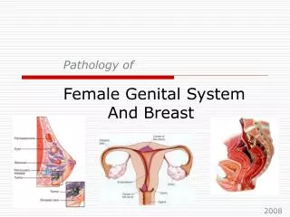
Management of patient with breast lump & nipple discharge. Objectives:. Anatomy of the breast Approach to a patient with breast lump Common breast problems (benign & malignant) Approach to a patient with nipple discharge. Anatomy of the breast. Anatomy of the breast :.

E N D
Objectives: • Anatomy of the breast • Approach to a patient with breast lump • Common breast problems (benign & malignant) • Approach to a patient with nipple discharge
Anatomy of the breast : • Modified sweat gland Extends from 2nd-6th rib & from sternal edge-midaxillary line. • Positioned over the muscles of the chest wall (the pectoralis major, serratus anterior, external oblique, and rectus abdominus fascia) • Attached to the chest wall by fibrous strands called Cooper’s ligaments ( suspensory ligament) which extend from the deep fascia beneath the breast and attach to the dermis of the skin. Carcinoma invading these ligaments may result in skin dimpling
Cont, anatomy of the breast • The breast is composed of glandular ducts and lobules, connective tissue, and fat. • The nipple and areola are separate structures. The unique anatomy explains why 18% of malignant cancers are found in the subareolar region • most breast cancer is thought to originate in the terminal ductal lobular unit (TDLU)functional secretory unit. • Half of this glandular tissue is located in the upper outer quadrant; therefore, nearly one half of all breast cancers occur in this area.
Blood supply : Venous drainage : -internal thoracic vein. -lateral thoracic vein. -Mainly the axillary vein. - intercostals veins . Arterial supply : - perforating branches of the internal thoracic artery(internal mammary artery)60%. - the lateral thoracic artery . - branches of the axillary artery. ( the thoraco-acromial artery& long thoracic artery ). - intercostal artery perforators . Most major venous pathways lead to the pulmonary capillary network (why lung metastases are common) or the vertebral veins (skeletal metastases).
Lymphatic drainage : • Interlobular lymphatic vessels sub areolar plexus (sappey’s plexus) (75%) of the drainage to the axillary lymph nodes. • Medial aspect of the breast internal mammery lymph nodes or the axillary lymph nodes.
How to manage a patient with breast lump or nipple discharge ?
History : Personal: Age gender Analysis of C/C SOCRATES 1- Pain 2- Lump 3- nipple discharge 4- abnormal appearance Previous Hx of breast problem Associated symptoms Constitutional symptoms Chronic illnesses Family Hx:
Cont’ History DDx: Hx of trauma to the breast Any medications ask about the risk factors of breast cancer : - Radiation exposure - Menstrual hx: Early menarche Late menopause and late pregnancy. Lactation Metastasis Hx - General malaise, weight loss - Recent backache, Bone ache - Jaundice - Mental changes - Dyspnoea, pleuritic pain - Nodules in the skin
Examination Examination: (A) Local Ex: - Position - Inspection - Palpation: (Feel, press, percussion, move, …. and surrounding tissues) - Lump: “4S”, “2T”, edge and composition - L.N. Axillary and supraclavicular (B) General Ex: Abdomen, lumbar spine … Points in Examination: Look for - Firm mass of variable shape and size - Fifty percent of masses found in the upper outer quadrant of the breast - May have associated pain with palpation, but most are painless - Nipple discharge or inversion - Skin retraction or tethering - Axillarylymphadenopathy - Inflammatory changes of the skin (e.g. peaud'orange)
DDx: Swelling of the whole breast: Bilateral -pregnancy, lactation - Idiopathic hypertrophy - Drug induced (e.g. cimetidine) Unilateral - Enlargement in the newborn - Puberty
Investigation • Imaging Studies • -Mammography • -US • -MRI • -Other imaging modalities • Diagnostic Procedures
Mamography special type of X-ray imaging used to create detailed images of the breast. The initial investigation for symptomatic breast in women older than 35 years. 95% accurate ( 5% - 10% ) false – ve. 2 views of each breast is taken as standard mammography: - 45° oblique. Mediolateral (MLO) - Craniocaudal position (CC) Additional views are obtained to clarify questionable lesion: -latero-medial (LM) -medio-lateral (ML) views -exaggerated CC views -magnification views -spot compression views -others Unreliable: because of high dense glandular tissue below the age of 35 years. Lactating lady .
Indication of mammogram : Screening : -Baseline mammogram for women ages 35-39 years. -Mammogram every 1-2 years for women ages 40-50 years. -every year once they reach 50 years of age . diagnostic: Metastatic adenocarcinomawithout known primary. Nipple discharge without palpable mass. Follow up
Ultra-sound - The most useful study in youngerwomen < 35 years with palpable breast mass. - Effective for lesions > 0.5cm. - Easily distinguishes cystic from a solid mass. Cystic: well defined, round, echo-free lesion with posterior enhancement. Solid: has echo within it & posterior enhancement. the introduction of Doppler enable definition of characteristic blood flow patterns. This can aid in separating benign and malignant lesions and distinguishing lymph node metastases from normal or reactive lymph nodes.
Fibroadenoma & Cyst in US : Fibroadenoma Cyst
MRI • Useful but expensive. • Usually used in screening of familial cases of breast cancer rather than X-ray which could be potentially harmful. • Distinguish scar from recurrence in women who have had previous breast conservative therapy for cancer (although it is not accurate within 9 months of radiotherapy because of abnormal enhancement). • The gold standard for imaging the breasts of women with implant.
CT scan CT is primarily used to evaluate for extramammary involvement of the tumor.
Fine needle aspiration (FNA) Indications:Establish cytological diagnosis. Advantages: Minimally invasive office procedure that is well tolerated by the patient. Often allows for a single trip to operating room. Specimen can be processed and interpreted rapidly. Disadvantages: 1- False (+) rate for cancer varies from 0%-1% on an institutional basis. 2- Significant false (-) rate ( >20%) for cancer because of small sampling size . Non palpable mass : stereotactic , Ultrasonographic or MRI- guided
Core-needle biopsy (CNB): Indications: Establishhistological diagnosis for < 3 cm mass. Advantages: -Minimally invasive, low-morbidity office procedure -False (+) rate for cancer is 0. Disadvantages: -Rare complications of hematoma and pneumothorax. -Significant false (-) rate (>20%)for cancer because of small sampling size. Non palpable mass :stereotactic (visualization by mammogram , Ultra-sonographic or MRI- guided CNB well tolerated &False (-) rate for cancer is approximately 1%.
Incisionalbiopsy:not used usually Indications: Establish histological diagnosis for a large mass (>3cm) when FNA and CNB are non-diagnostic. Advantages: -Performed under local anaesthesia. -False (+) rate for cancer is 0. -False (-) rate for cancer is close to 0. Disadvantages: -Substantially higher cost than FNA or CNB. -Open Surgical procedure with associated risks of bleeding and wound infection.
4.Excisional biopsy: Indications: Establish definitive histological diagnosis for a small (<3cm) mass when FNA and CNB are non-diagnostic. Advantages: • Can be therapeutic as well as diagnostic for benign mass and for malignant mass excised with negative microscopic margins. • False (+) and false (-) rates for cancer are 0. • Performed under local anaesthesia. Disadvantages: • Open Surgical procedure with risks of bleeding and wound infection. • Substantially higher cost than other biopsy procedures.
Wire localization breast biopsy:for non-palbable mass Indications: Establish definitive histological diagnosis for a non-palpable but visualized abnormality. Advantages: -Therapeutic as well as diagnostic for benign masses and for malignant masses excised with negative margins. -False (+) rate for cancer is 0. -False (-) rate is 0 if visualized abnormality is completely excised. Disadvantages: Open procedure that requires radiological localization before surgical excision. Occasional (1%) failure to excise abnormality. May require relocalization and reoperation. Cosmetic deformity may result.
common causes of a benign breast mass Fibrocystic disease: the most common breast mass in women. Fibroadenoma: the most common benign tumor. Fat necrosis Abscess Cyst Others : - Intraductalpapilloma - Ductal/ lobular Hyperplasia - Ductectasia - Lipoma - Granulomatous mastitis Note :1- in general, Mass: cystic or solid, Tumor: solid 2- the difference between fibrocystic changes and fibroadenoma is that in fibrocystic changes u cant define a mass while fibroadenoam is a mass
Fibrocystic change: Benign changes - Age:30 – menopause (and after if HRT used) - C./F. : Breast pain, swelling, with focal area of nodularity, freq. bilateral, mobile and varies with menstrual cycle… - No increase risk of breast cancer but makes evaluation of mammographic malignant changes more difficult. • Treatment: If >40 y : mammography every 3 years analgesia, OCP or danazol for sever symptoms.
Fibroadenoma: - Most common benign breast tumor in women < 30y - No malignant potential except if sclerosingadenosis present. - C./F. nodules: smooth, rubbery, discrete, well-circumscibed, non-tender, mobile, hormone dependent . - Unlike cysts, needle aspiration yield no fluid Investigations: - Mammogram - US - FNA to R/O solid lesion Rx: - Generally conservative – serial observation -Excision if mass rapidly growing, if >5cm in size or if Pt. wants , equivocal result , if the pt has no access for follow up, if there is family history of cancer.
Phylloid tumor (cystosarcoma): • Rare type of fibroadenoma. • typically large, fast growing masses that form from the periductalstromal cells of the breast. • most common between the ages of 40 and 50, prior to the menopause. • Although it is mostly benign , It can recur after excision . • The malignant form (10%) can metastasize hematogenously most commonly to the lungs . • The common treatment for phyllodes is wide local excision.
Fat necrosis - Result of trauma (may be minor, +ve trauma Hx in only 50%) - Firm, ill-defined mass with skin or nipple retraction +/- tenderness • Regress spontaneously, but complete excisional biopsy to rule out carcinoma . • It resembles cancer clinically & radiologically. The only way to differentiate is by biopsy.
Abscess: - Unilateral localized pain and erythema. - R/O inflammatory carcinoma, as indicated - Staphylococcus aureus are the most common organisms .
Cyst: C\F :Fluid-filled sacs that often feel like soft grapes. Can sometimes be tender, especially just before the menstrual period. • Cysts may be drained in the clinic. Rx: • If the fluid removed is clear or greenish, and the lump disappears completely after it is drained, no further treatment is needed. -If the fluid is bloody, it is sent to the lab to look for cancer cells. If the lump doesn't disappear, or recurs, it is usually removed surgically.
Galactocele : • is a cystic tumor containing milk or a milky substance that is usually located in the mammary glands. • Galactoceles are benign and are not a cause for concern. • It is caused by a protein plug that blocks off the outlet. Once lactation has ended the cyst will resolve on its own without intervention. • A galactocele does not cause infection as the milk within is sterile and has no outlet for which to become contaminated. • Attempts to drain the cyst are unsuccessful because the protein plug remains intact and milk production continues.
Granulomatous mastitis: • Characteristic for granulomatous mastitis are multinucleated giant cells and epithelioidhistiocytes around lobules. Often minor ductal and periductal inflammation is present. The lesion is in some cases very difficult to distinguish from breast cancer. • most often completely aseptic but infectious causes must be considered as well. • C\F:distinct firm mass mostly in the subareolar region. • PREDISPOSING FACTORS: -2 years and up to 6 years after pregnancy, usual age range is 17 to 42 years. -Use of hormonal contraceptives, prolactin raising medications and hyperprolactinemia .
Epidemiology: • The 2nd leading cause of cancer mortality in women (1st?) • - Lifetime risk : 11-13%
Risk factors of breast cancer: • - 99% female • - 80% >40 y.o. • - Prior Hx of BC, prior breast biopsy. • 1st degree relative with BC( incr. risk if premenopausal ) • risk in (HYPERESTROGENEMIA STATE): - early menarche <12y - late menopause>55y - 1st pregnancy >30y, - nulliparity - OCP - HRT for 5y • - risk with lactation, early menopause, early childbirth • - Radiation exposure • - Hx of specific benign breast disease ( Atypical hyperplsia 4x )
1- Non- invasive: a) Ductal carcinoma in situ (DCIS) - Completely contained within breast ducts - 80% non-palpable, detected by screening mammogram b) Lobular carcinoma in situ (LCIS) -Completely contained within breast lobule -No palpable mass, no mammographic findings, usually incidental finding on breast biopsy.
2- invasive: • Infiltrating ductal carcinoma (most common 80%): hard ,scirrhousthe most common type ,infiltrating tentacles Papillary ,medullary ,mucinouse ,tubular cancers Generally better prognosis. • Invasive lobular carcinoma (8-15%): -20% bilatral -Dose not form microcalcification , harder to detect mammographically . • Paget’s disease (1-3%): Ductal carcinoma that invades nipple with scaling ,eczematous lesion .
Inflammatory carcinoma (1-4%) Ductal carcinoma that invades dermal lymphatics Most aggressive form of breast cancer Erythema , skin edema ,warm, swollon ,tender +- lump • Male breast cancer (<1%) Most commonly infiltrating ductal carcinoma Often diagnosed at later stages • Sarcoma Rare ,most commonly cystosarcomaphyllodes , a variant of fibroadenoma • Lymphoma –rare