Download

1 / 33
1.1k likes | 4.04k Vues
Care of the Patient with an EVD or Lumbar Drain. Lauren Walker RN, BSN, CCRN. CSF and Ventricular System. CSF is a colorless, clear fluid produced by choroid plexus in lateral and third ventricle Functions as a cushion for brain and spinal cord 500-600 ml CSF produced daily

E N D
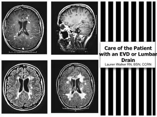
Care of the Patient with an EVD or Lumbar Drain Lauren Walker RN, BSN, CCRN
CSF and Ventricular System • CSF is a colorless, clear fluid produced by choroid plexus in lateral and third ventricle • Functions as a cushion for brain and spinal cord • 500-600 ml CSF produced daily • 3 cycles of CSF, made then absorbed, made then absorbed, etc in a 24hr period • Brain has four CSF filled and interconnected ventricles
Intracranial Pressure • Intracranial Pressure (ICP) is the pressure exerted by the brain content: • Brain Tissue (85%) • Cerebral Spinal Fluid (CSF) (10%) • Intravascular blood (2-11%) • Cerebral Autoregulation: The ability of the cerebral vessels to constrict and dilate as needed to maintain adequate cerebral perfusion • Impaired with brain injury and the cerebral blood flow becomes passively dependent on blood pressure • Monro-Kellie Hypothesis: an increase in one component must be offset by an equal decrease in one or more components otherwise an increase in ICP will result
ICP Pressures We want ICP to be less than 20! • Increased ICP occurs when the intracranial volume exceeds the brain’s ability to compensate for increased volume • Sustained ICP above 25 mmHg is considered a neurologic emergency
Cerebral Perfusion Pressure • Cerebral Perfusion Pressure (CPP):Pressure at which the brain is perfused • About 15-20% of cardiac output CPP = Mean Arterial Pressure (MAP) – ICP. • Normal CPP 40-60 (Kids, depends on age) • Normal CPP 80-90 mmHG (Adults) • CPP below set parameter will result in cerebral ischemia and tissue death
Contusions Hematomas Tumors Infarcts Fluid overload Mechanical ventilation (PEEP) Valsalva maneuver Coughing Endotrachial suctioning Hypercarbia Hypoxia Increased CSF Production Metabolic disturbances Causes of Increased ICP
ICP Monitoring and the EVD • Intraventricular catheters are the gold standard for measuring ICP • Placed directly in the ventricle (typically in the anterior horn of the lateral ventricle through a burr hole in the skull) • Attached to a pressure transducer • An external ventricular device (EVD) • Has ICP monitoring capabilities • Can also assist with controlling increased ICP by CSF drainage • The main disadvantage to an EVD is that it is the most invasive device because it penetrates the meninges and brain
Expected Outcomes of ICP Monitoring and CSF Drainage • Avoid secondary injury • Patient will maintain adequate CPP • WBC, CSF cell count, protein, glucose and culture will be within expected limits • Patient/family will receive education! • Visitors will verbalize understanding of limiting movement of patient
Nursing Management and Assessment Maintain optimal cerebral tissue perfusion • Perform and document neuro assessment every hour and PRN and note trends. Notify physician for changes in neurostatus, ICP, and CPP for more than 5 minutes. • Monitor Hourly: • Vital Signs: BP, pulse, respiration and temperature. • Pupil size and reaction • Glasgow coma scale • ICP • MAP • CPP = MAP – ICP
Nursing Management and Assessment Monitor the patient every hour and as necessary for symptoms of increasing ICP • Change in level of consciousness: • Headache • Restlessness/agitation • Nausea/Vomiting • Seizure activity • Visual changes • Changes in LOC • Changes in pupillary responses • Labored respirations and/or pattern • Changes in motor strength • Cushing’s Triad: Increasing BP with a widening pulse pressure, decreased HR, irregular respirations
Nursing Management and Assessment Assessment of the EVD • Inspecting EVD from insertion site along the entire drainage system • Check for cracks in the system or fluid leaking from the insertion site • Dressing is intact • Hourly assessment of CSF drainage amount, color, and clarity. • Ensure the system is appropriately clamped or open depending on order. • Check patient position to ensure transducer is at the ordered reference level. • If the patient is very active and moving around in bed, it is imperative to frequently assess that the drain is leveled appropriately to prevent over- or under-drainage.
Nursing Management and Assessment Facilitate venous return: • Patient’s head may be elevated according to the discretion of the attending and neurosurgeon. • The head position must remain neutral, may use soft cervical collar or sandbags, if necessary. • Instruct family members regarding the plan of care and the need to limit stimulation of patient r/t head injury.
Nursing Management and Assessment • Identify activities that alter intracranial pressure • lights, noise, repositioning • Arrange nursing care to minimize elevation in ICP (cluster care) • Assess ICP waveform continuously • Body temperature should remain below 38 C. • Assure prescribed seizure prophylaxis is instituted if indicated.
Nursing Management and Assessment • Monitor labs, as ordered. • Maintain HCT greater than 25%. • If patient is receiving Mannitol, obtain Serum Osmolarity every 6 hours. (Notify MD prior to administering if serum osmo > than 310. • Record I/O every hour. • If urine output is greater than 200 mL for 2 hours and specific gravity is less than 1.005, not associated with Mannitol dosing, notify neurosurgeon
EVD Complications • An absence of an ICP waveform • May be the result of air bubbles, clots, or debris within the drainage tubing or across the transducer. • A malfunctioning pressure cable, module, or transducer may also result in the loss of the ICP waveform • Infections • Manipulation and accessing of drainage tubing can be a source of contamination • Drainage tubing accidentally becomes disconnected: every effort should be made to maintain the sterility of the ventricular catheter. • New sterile EVD tubing should be obtained and connected
EVD Complications • CSF Overdrainage • Maintain the drip chamber at the prescribed zero reference and pressure levels • Family and Pt education!! • changing the bed position is to be performed only with assistance. • Raise the drain any time there is a patient response or procedure that may cause CSFoverdrainage • Clamp for coughing, vomiting, suctioning, or repositioning.
Leveling EVD • The transducer on the set-up is leveled to the patient’s external auditory meatus. • The “pressure level” indicator (located on the drip chamber of the collection system) is aligned to the ordered cm H2O above the external auditory meatus. • If continuously draining CSF, turn the drain off to the patient momentarily to obtain the ICP reading.
Zeroing the TransducerEVD • Raise or lower the system measured to the tragus of the ear • Lower the Buretrol to the “zero point” on the system by pinching the tabs together above the buretrol and lowering into alignment with zero on the device • Turn the stopcock “off” to the patient (open to the Buretrol/transducer) • Do Not open the transducer to air (the transducer will be opened to air automatically through the vent on top of the Buretrol). • Highlight the ICP box on the GE monitor screen and select “zero ICP” on the drop down menu choices. Wait for a “0” to appear on the monitor for the ICP reading. • Replace the Buretrol to the position on the pole mount that has been ordered by the physician. • Zero the ICP every 12 hours
Obtaining an ICP Tracing • Turn stopcock “off” to the drain and “open” to the transducer to obtain an accurate ICP numerical value and waveform. • The ICP numerical value and waveform should be obtained every hour. • If there is an increase in intracranial pressure, then the value should be obtained more often (i.e., Q15 minutes).
Draining CSF • For Continuous ICP Measurements: • Keep the EVD stopcock “off” to drain and “open” to the transducer for continuous ICP monitoring. • If ordered by MD, when the ICP reaches a specified pressure, “open” the stopcock to drain CSF for a short time period. • Continuous CSF Drainage/Intermittent ICP Monitoring • The EVD stopcock is “off” to transducer • The amount of CSF drainage is controlled by raising the pressure level on the graduated burette above the Foramen of Monro, which is the zero reference level. The EVD device does not allow practitioner’s to drain CSF and monitor ICP simultaneously.
Changing the Drainage Bag • The collection bag is changed when about three quarters full. • Wear sterile gloves and a mask. • Cleanse the stopcock below Buretrol with Betadine (Scrub connection for 30 seconds). • Assure the stopcock between the Buretrol and the collection bag is “off” toward Buretrol. • Using sterile handling technique, disconnect the drainage collection bag from the Buretrol stopcock and detach bag from the system panel. (Handing the full bag off to a second person is recommended.) • Connect a sterile replacement collection bag to Buretrol stopcock and attach to system panel. • Cap off the open port of the full collection bag and dispose in red trash bag.
Patient Transport • Clamp the EVD during transport, when repositioning or with any patient movement that would cause the zero point of the EVD/LD to be lower than the reference point. • Re-zero, level and re-open the EVD/LD after patient transport or movement is completed.
ICP Monitoring and Lumbar Drains • Lumbar drainage devices (LD) are closed sterile systems that allow the drainage of CSF from the subarachnoid space. • LD are inserted via a specialized spinal needle into the lumbar subarachnoid space at the L2–L3 level or below • avoids injury to the spinal cord • In the lumbar CSF space, the spinal catheter will be alongside the cauda equina • Consists of the ventral and dorsal spinal nerve roots that descend from the spinal cord and exit the spinal canal at lumbosacral levels
Lumbar Space ←Dural Tube L1 pedicle→ ←Conus Medularis L2 pedicle→ ← Cauda Equnia Spinal Cord ends at L1-L2 at Conus Medularis LD is inserted in lumbar subarachnoid space: L2 to L3 space avoiding injury to spinal cord Sacrum→
Indications for a Lumbar Device • Postoperative or traumatic dural fistula (CSF leak) • Treatment of shunt infections • Diagnostic evaluation of idiopathic normal pressure hydrocephalus • Reduce ICP during a craniotomy • Adjuvant therapy in the management of TBI • Thoracoabdominal AAA to improve spinal cord perfusion • Manage nontraumatic subarachnoid hemorrhage to prevent vasospasm • Manage increased ICP associated with cryptococcal meningitis
Patient Care with LD • Patient positioning is crucial to prevent complications • The head of the bed, height of drainage chamber, and changes in patient positioning must be monitored closely to prevent sudden overdrainage. • Patients may turn from side to side without significant impact on drainage unless the catheter is found to be positional. • Nursing staff should monitor which positions result in variances in drainage rate from the LD and plan patient care • Perform a complete motor and sensory exam with neuro exam • Assess for s/s of meningeal irritation • Photophobia, neck stiffness, N/V, HA, bowel and bladder function
Lumbar Drain Complications • Overdrainage • Movement and repositioning of the patient • Not frequently observing the amount draining • Infection at insertion site • Manipulation of catheter and tube could be a source of contamination • Non-occlusive dressing • Tube Dislodgement • Lower extremity weakness • Changes in bowel and bladder patterns • Could indicate a nicked nerve • Tracking in tubing • Bleeding in spinal cord
Lumbar DrainZeroing • There is no such thing • We are not measuring ICP with a Lumbar Drain • No transducer on system • No need for zeroing! (One less thing for you to do!)
LD Leveling • The transducer on the set-up is leveled to the patient’s shoulder or iliac crest (per NP/MD order) • The “pressure level” indicator (located on the drip chamber of the collection system) is aligned to the level at which it drains the order volume Iliac Crest↓
LD Drainage • 1. Draining to a specific volume: MD orders a specific amount of CSF to be drained at a particular time period • Manipulation of the drain to achieve specific amount • 2. Draining at a specific level: MD orders level of the pt’s body • Continuous drainage (ICU monitoring) • 3. Draining at a specific pressure: MD orders a specific pressure for draining • System drains only when the pressure exceeds the preset pressure • Usually for shunt infections/malfunction • ICU Monitoring
References American Association of Neurosciences Nurses. (2011). Care of the patient undergoing intracranial pressure Monitoring/External ventricular drainage or lumbar drainage. AAN clinical practice guideline series. Glenview, IL: Codman and Shurleff. http://www.aann.org/pubs/content/guidelines.html. Barker, E. (Ed.). (2008). Neuroscience nursing, A spectrum of care (3rd ed.). St Louis, MO.: Mosby Elsevier. Buchbinder, D. (2012). AO Surgical Reference. Retrieved April 18, 2012, from https://www2.aofoundation.org/wps/portal/!ut/p/c0/04_SB8K8xLLM9MSSzPy8xBz9CP0os3hng7BARydDRwML1yBXAyMvYz8zEwNPQwN3A_2CbEdFAEfu4iM!/?showPage=rehabilitation&bone=CMF&segment=Cranium&classification=93-Skull+base,+Temporal+bone+(lateral+skull+base)&treatment=&method=Delayed+management+of+hearing+loss&implantstype=&approach=&redfix_url=1289912913263. Inova Health System. (2011). Critical care standard 2.400 intracranial pressure monitoring- procedure and protocol. Unpublished manuscript. Wiegand, D. (Ed.). (2011). AANC procedure manual for critical care (6th ed.). St. Louis, MI: Elseview Saunders.