Download
1 / 93
930 likes | 1.16k Vues
" Emergency Anaesthesia for the Head Injured Patient for non head injury procedures". Dr. J. Balavenkat MD, DA Consultant Anaesthesiologist Ganga Hospital Coimbatore. Man kills Man … On the Roads!. Global Road Traffic Injury-Related Mortality.

E N D
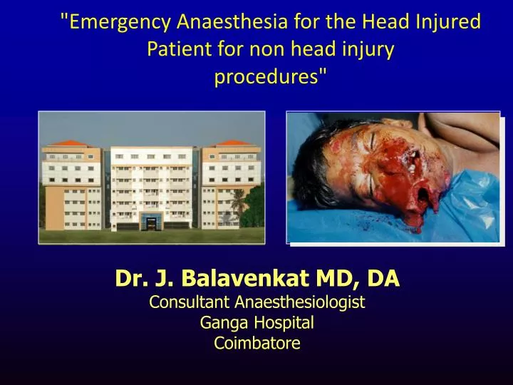
"Emergency Anaesthesia for the Head Injured Patient for non head injury procedures" Dr. J. Balavenkat MD, DA Consultant Anaesthesiologist Ganga Hospital Coimbatore
By 2050, India will have the greatest number of automobiles on the planet, overtaking the United States
Before we coclude this two day meet about 500 people would have died and more than 5000 injured in India
If you are between 15-45 years of age you have more chance of dying due to an accident than any other disease
Statistics • Nearly 1.5 to 2 million persons injured every year in India • Road traffic injuries are the leading cause (60%) of TBIs followed by falls (20-25%) and violence (10%) • Main cause of death for patients who survive >48 hours
Initial Resuscitation and Investigation protocol? • CT Scan : Timing in an unstable patient
Bleeding is a major cause of death in trauma* N=289 * Patients dying in hospital within 48 hours Sauaia A et al. J Trauma 1995;38:185-93
Contentious Issues….. • Role of hyperventilation • Role of hypothermia • Role of Glucose level in the neurological outcome • Best fluid to resuscitate • Cerebral protective strategies.
Pathophysiology of TBI is complex.But you do not need to know the details Simple, early and focussed interventions are effective in preventing secondary injury
Basic pathophysiology of brain injury • Primary insult: • Direct tissue damage at the time of impact • What are causes of secondary brain injury? • Tissue injury occurring after the initial injury due to multiple causes • Raised ICP, hypotension, hypoxia, hypo and hypercarbia, hyperthermia,hypo and hyperglycaemia, seizures • Treatment goal: limit or prevent these secondary insults
GLASGOW COMA SCALE SCORE EYE OPENING 4 Spontaneous 3 To speech 2 To pain 1 None BEST VERBAL 5 Orientated 4 Confused 3 Inappropriate words 2 Incomprehensible sounds 1 None BEST MOTOR 6 Obeys commands 5 Localises to pain 4 Withdraws to pain 3 Abnormal flexion to pain 2 Extension to pain 1 None
Key issue is prevention of secondary injury • Attempt to maintain cerebral blood flow (CBF) • Attempt to maintain cerebral oxygenation • Applicable in all areas • Pre-hospital • ED • Peri-operative • Intensive Care
Immediate management • A, B, C approach • Airway management must be with C-spine immobilisation and control • If GCS ≤ 8 should be intubated and ventilated to low normocapnia [4-4.5 kPa, 30-35 mmHg] • A rapid sequence induction/intubation is appropriate, but avoid ICP ↑ • Circulation must be supported with IV volume loading and a MAP of 90mmHg maintained
Intubation of the patient with Brain Injury • Record GCS and pupils before RSI • Rapid Sequence oro-tracheal intubation: • In line stabilization of C-spine • Pre-oxygenation with 100% O2 • Induction agent? • NMB: suxamethonium or rocuronium? • OGT or NGT?
Actions after tracheal intubation? • Confirm TT and gastric tube placement (clinical and CXR) • Ventilate, aiming for PaCO2 4-4.5 kPa (30-35 mmHg) • Oxygenate, using PEEP if necessary • PaO2 > 13 kPa (97 mmHg) SaO2 >94% • Cardiovascular assessment and stabilization • IV volume • MAP ≥ 70-90 mmHg
Circulation Hypotensive bleeding patients • Early Vs Delayed Fluid Resuscitation • Large Vs Small Volume resuscitation • Crystalloids or colloids? • To Transfuse blood and blood products early or late?
Circulation CLASSIFICATION OF HYPOVOLEMIA
Goals of Resuscitation before controlling haemorrhage in a head injured • Systolic BP of 100 mmhg • Heart rate <120 • Hb > 9.0 gms/dl • Urine output >0.5 ml\kg\hr • SpO2 > 96 • Serum Lactate less than 2.2 mmol/lt
Third generation HES: Tetrastarch • Positive effect on tissue oxygenation • Improves microcirculation • Less effect on coagulation • No effect on liver function • Even with 50ml/kg body wt renal parameters not altered • Tissue accumulation less.
Acute extradural midline shift ventricle compression
Acute subdural Compression and shift
What are indications for head CT? • GCS less than 13 at any point since the injury. • GCS equal to 13 or 14 at two hours after the injury. • Suspected open or depressed skull fracture. • Signs of basal skull fracture (haemotympanum, ‘panda’ eyes, cerebrospinal fluid otorrhoea, Battle’s sign). • Post traumatic seizure. • Focal neurological deficit. • More than one episode of vomiting • Amnesia for >30 minutes of events before impact
Transfer from secondary to tertiary care settings • Prevent further injury during transfer to tertiary care • Agree transfer guidelines between the referring hospital and the neurosurgical unit • Resuscitate and stabilise the patient before transfer • beware the undiagnosed abdominal injury • All patients with a GCS <9 (and maybe higher) requiring transfer to tertiary care should be intubated and ventilated
Basic Treatment • A: Tracheal tube • B: Ventilate to pCO2 4-4.5 kPa (30-35 mmHg), pO2 ≥ 13 kPa (95 mmHg) • C: MAP ≥ 70 mmHg (aim CPP 50-70 mmHg) • IV volume • PEEP is allowed • Arterial and CVP lines • Vasopressors: noradrenaline initially • D: Monitoring • Pupils • Blood glucose
Remember • Hypoxia: 50% of patients with severe head injury have PaO2< 10 kPa (75 mmHg) • Mortality is increased by 20% if hypoxia present on admission • MAP < 90 mmHg = 30% increase in mortality • Metabolic: Glucose > 10mmol/L (180mg%) = worse
Cerebral Perfusion Pressure • CPP = MAP – ICP • Normal CPP = 70 – 100 mmHg • Normal ICP = 0 – 15 mmHg • Evidence supports favorable outcome with CPP aim of 50-70 mmHg • Initially CPP is not known • Maintain MAP >70 mmHg in an attempt to maximize CPP
Acute treatment of increased ICP • Check A, B still OK • beware partially blocked ETT or chest wheeze • Cautious, short-lived, hyper-ventilation • Maintenance of circulation and CPP • Osmotherapy • Neurosurgical interventions • Decompression • CSF removal
Hyperventilation • Theory: ICP via constriction of cerebral vasculature and reduced brain volume • Change: 2- 4% in cerebral blood flow with every 1mmHg change in pCO2 • PaO2 below 60 mmHg (8 kPa) is the second most powerful predictor of poor outcome • PaCO2 <4 kPa (30 mmHg) may increase ischaemic damage due to cerebral vasoconstriction • Severe vasoconstriction with pCO2 < 25mmHg (3.5 kPa) • Evidence suggest hyperventilation is harmful
Hyperventilation: Recommendations • Brain Trauma Foundation: 1) Prophylactic hyperventilation is to be avoided 2) Hyperventilate for brief periods when there is acute neurologic deterioration 3) Hyperventilate for ICP that is refractory to sedation, paralysis, cerebral spinal fluid (CSF) drainage, and osmotic diuretics
Osmotherapy • Mannitol • Hypertonic saline
Mannitol • Lowers ICP and Increases MAP • 1) plasma expander: increases cerebral blood flow and cerebral oxygen delivery • 2) Delayed effect (30 minute - 6 hour): osmotic agent • Cardiovascular collapse if volume depleted • May increase bleeding • Renal failure: if serum osmolarity > 320 mOsm • Concentrated in brain tissue with prolonged infusion
Mannitol: Recommendations • Brain Trauma Foundation: 1) No standard for mannitol 2) Effective for control of ICP: intermittent boluses more effective than continuous infusion. (dose: 0.25 g/kg to 1 g/kg) 3) Indications for mannitol prior to ICP monitoring: signs of transtentorial herniation or progressive neurological deterioration 4) Euvolemia should be maintained: fluid replacement • No role for high dose
Intravenous Fluids: Hypertonic Saline • Hypertonic Saline: 5% or 7.5% NaCl • Increases MAP and reduces ICP • Hypothesis: cerebral swelling may be prevented by altering the osmolar load • Keep serum Na < 155 mmol/L [some centres will accept ~ 170] • No central pontine myelinolysis reported as this is not rapid correction of hyponatraemia
Intravenous Fluids • 0.9% saline may be better than Ringers lactate • Hematocrit: • Level of 30-33% believed optimal • < 30% exacerbates neurologic injury • Glucose: • Hyperglycemia increases neurologic injury • Prevent hypoglycemia • Use of insulin improves outcome in lab animals • Goal: blood sugar 100 – 150 mg/dl (~5-8 mmol/l)
Barbiturates • ICP by suppressing cerebral metabolism • No evidence that this is associated with reductions in mortality or disability. • Occurrence of hypotension • Significant fall in body temperature
Barbiturates: current recommendations • Intractable ICP not responding to sedation • Burst suppression on EEG • Thiopentone drug of choice • Caution: needs multimodality monitoring
Associated Injuries….. • Spinal Cord Injury • Faciomaxillary Injury • Thoracic Injury • Abdominal Injury • Pelvic Injury • Musculoskeletal Injury
Spinal Cord Injury • Hypotension (neurogenic shock) – characterized by bradycardia rather than tachycardia (due to loss of cardiac accelerator function and unopposed parasympathetic tone) may be difficult to distinguish from hypotension due to acute hemorrhage. • The hypotensive trauma patient is always assumed to be bleeding, until this possibility is definitively ruled out.
Early Need for a Surgical Airway….. • Head injury/coma • Face or neck injury • Cervical Spine injury • Chest injury: rib fractures • Haemo-pneumothorax
Rib Fractures • Ribs 1-3 major force to break :associated injuries • Ribs 4-9 Lung contusions : Haemothorax/pneumothorax • Ribs 10-12 Intra-abdominal injuries
What are the causes of life threatening chest trauma? Primary survey • Airway obstruction • Tension pneumothorax • Open pneumothorax • Massive haemothorax • Flail chest • Cardiac tamponade