BAKTERI
BAKTERI. OLEH SUDRAJAT FMIPA UNMUL 2009. Klasifikasi Ba k teri. Gram + cocci Gram - bacilli. Klasifikasi Ba k teri. Gram - Spirochete Gram + bacilli. Microscopic Phenotypic Exam. Gram positive. Gram stain distinguishes between Gram + and Gram – bacteria


BAKTERI
E N D
Presentation Transcript
BAKTERI OLEH SUDRAJAT FMIPA UNMUL 2009
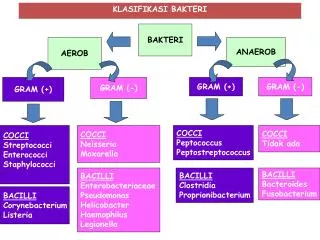
Klasifikasi Bakteri Gram + cocci Gram - bacilli
Klasifikasi Bakteri Gram - SpirocheteGram + bacilli
Microscopic Phenotypic Exam Gram positive • Gram stain • distinguishes between Gram + and Gram – bacteria • narrows the possibilities quickly Gram negative
Microscopic Phenotypic Exam • special stain • allows for the distinction of microorganisms with unique characteristics • capsule • acid fast staining detects the waxy presence of Mycobacterium tuberculosis Capsule staining Acid fast staining of M. tuberculosis
Staining Organisms • needed to allow us to see the organisms using light microscopy • organisms are killed in the process • Simple stains • stain is applied and colours the organism e.g. methylene blue
Complex Stains • stains may be combined which stain different structures different colours. e.g. giemsa stains malarial parasites nucleus red and cytoplasm blue • stains may be applied in sequence with a step to remove stain in between. e.g. gram stain - a key stain in microbiology!!
The Gram Stain • Developed by Christian Gram in the 19th Century • He found that a stain could be washed out of some organisms much more easily than others • Technique allows differentiation of many bacteria into 2 groups: gram positive and gram negative – corresponding to cell wall type. • Continues to be used extensively and is important!
Method for Gram Stain • Crystal violet – stains all the bacteria dark purple • Iodine – binds to crystal violet and fixes it (acts as a mordant) • Alcohol/Acetone washes out the stain from gram negative bacteria • (Gram originally stopped here, so that organisms that stained purple were “positive” because they could be seen; subsequently the fourth step was added so that both the positive and the negative organisms could be seen.) • Safranin stains the gram negative bacteria pink.
Acid Fast Stain • Some bacteria cannot be stained by the gram stain because of lipids in the cell walls. (e.g. Mycobacterium tuberculosis, the tuberculosis bacterium) These bacteria may be stained by an “acid fast method”. • involves: - staining with a strong red stain (to “force” the stain in ) • washing out the stain with a mixture of acid and alcohol • restaining (“counterstaining”) with a blue or green stain. • Acid Fast organisms are Red. These are sometimes called AFB (acid fast bacilli). • Other organisms are the colour of the counter stain (blue or green).
Ciri-ciri bakteri gram negatif Dinding sel tipis (10-15 nm) berlapis tiga (multi). Kandungan lipid tinggi : peptidoglikan (10% berat kering), tidak ada asam tekoat. Kerentanan terhadap penisilin kurang rentan. Pertumbuhan tidak begitu dihambat oleh zat warna dasar.
Ciri-ciri bakteri gram negatif Persyaratan nutrisi → relatif sederhana. Resistensi terhadap gangguan fisik→ kurang resisten Kehilangan kompleks warna ungu kristal pada waktu dicuci alkohol → terwarnai pewarna tandingan safranin (sel tampak merah muda).
Contoh. Treponema pallidum merupakan salah satu bakteri spirochaeta. Bakteri ini berbentuk spiral. Terdapat empat subspecies yang sudah ditemukan, yaitu Treponema pallidum pallidum, Treponema pallidum pertenue, Treponema pallidum carateum, dan Treponema pallidum endemicum.. Treponema pallidum pallidum yang merupakan penyebab sifilis.
Sifilis atau penyakit Raja Singa adalah salah satu penyakit menular seksual (PMS) yang kompleks, disebabkan oleh infeksi bakteri Treponema pallidum. Perjalanan penyakit ini cenderung kronis dan bersifat sistemik. Hampir semua alat tubuh dapat diserang, termasuk sistem kardiovaskuler dan saraf. Selain itu wanita hamil yang menderita sifilis dapat menularkan penyakitnya ke janin sehingga menyebabkan sifilis kongenital yang dapat menyababkan kelainan bawaan atau bahkan kematian. Jika cepat terdeteksi dan diobati, sifilis dapat disembuhkan dengan antibiotika. Tetapi jika tidak diobati, sifilis dapat berkembang ke fase selanjutnya dan meluas ke bagian tubuh lain di luar alat kelamin.
Ciri – Ciri bakteri gram positif Struktur dinding sel tebal (15 – 80 nm) dan berlapis tunggal. Komposisi kimiawi : kandungan lipid rendah (1 - 4 %), peptidoglikan lapis tunggal (>50%), asam tekoat. Kerentanan terhadap penisilin→ lebih rentan (peka). Pertumbuhan dihambat oleh zat-zat warna dasar (misal ungu kristal)
Ciri - ciri bakteri gram positif Persyarataan nutrisi → relatif rumit pada banyak spesies. Resistensi terhadap gangguan fisik→ lebih resisten (tahan). Reaksi terhadap pewarna primer atau ungu kristal iodium → dapat menahan sampai akhir prosedur (sel tampak biru gelap/ungu).