Download
1 / 39
410 likes | 723 Vues
F211 Exchange and Transport Lungs. What do animals need to gain from their environment to stay alive? How do these substances get to cells? What do plants need to gain from their environment to stay alive? How do these substances get to cells?

E N D
F211Exchange and Transport Lungs What do animals need to gain from their environment to stay alive? How do these substances get to cells? What do plants need to gain from their environment to stay alive? How do these substances get to cells? Give 2 examples of wastes that living organisms have to get rid of somehow. How does the waste get from the cells to where it is excreted?
Learning Outcome You should be able to: • Explain, in terms of surface-area-to-volume ratio, why multicellular organisms need specialised exchange surfaces and single-celled organisms do not.
Increasing size and complexity • Single celled organisms do not need complex exchange and transport systems. • Why not? • How does exchange of substances take place? How do they transport substances into the centre? • Bigger organisms with several or many layers of cells and which are more active need specialised exchange and transport systems, why?
Complete the table to show how surface area to volume ratio changes as size of cube increases
Completed table to show how surface area to volume ratio changes as size of cube increases The bigger the object the less outer surface it has compared to its volume. This means it is harder for substances to move into the object and through to the centre of the object by diffusion.
Features of Exchange Surfaces? • Large surface area, often folded • Thin barrier to reduce diffusion distance • Fresh supply of required molecules on one side to keep concentration high • Removal of required molecules on other side to keep concentration low • (maintains concentration gradient)
Examples of exchange/ absorption surfaces • Alveoli, exchange of oxygen and carbon dioxide • Small intestine, absorption of nutrients • Liver cells (hepatocytes), absorption of metabolically active substances, blood sugar levels adjusted • Root hairs, water and minerals absorbed • Fungal hyphae, absorption of nutrients
Learning OutcomesYou should be able to: • Describe the features of an efficient exchange surface with reference to diffusion of oxygen and carbon dioxide across an alveolus. • Describe the features of the mammalian lung that adapt it to efficient gas exchange.
The Lungs and Associated Structures (familiar from KS3 and KS4)
The Lungs and Associated Structures (familiar from KS3 and KS4)
Features of the mammalian lung that enable efficient gas exchange • Individual alveoli are only100-300 micrometers across, very numerous ( about 300-500 million) total surface area = approx 70 m2 • Alveoli walls are one cell thick, plasma membranes surround a very thin layer of cytoplasm. • Capillary wall is only one cell thick • Cells are squamous, flattened • Capillaries in close contact with alveolus wall • Capillaries very narrow so RBCs are squeezed close to the walls and so close to the air in the alveoli • Total diffusion distance from inside alveolus to inside RBC is only about 1 micrometer • Surfactant stops alveoli collapsing due to cohesion of water when air pressure is low • Ventilation and blood transport maintains concentration gradients of oxygen and carbon dioxide for efficient gas exchange
Outline the mechanism of breathing (inspiration and expiration) in mammals, with reference to the function of the rib cage, intercostal muscles and diaphragm. Remember to relate air movements to changes in volume of chest cavity and hence changes in air pressure.
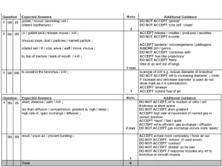
Describe the distribution of cartilage, ciliated epithelium, goblet cells, smooth muscle and elastic fibres in the trachea, bronchi, bronchioles and alveoli of the mammalian gaseous exchange system. What is the role of each of these tissues? (What is the definition of a tissue?) Make notes in the table supplied using pages 48 and 49 of your text book NB the role of surfactant (not a tissue) on the internal surfaces of the alveoli
Bronchiole and Trachea in transverse section Later in Unit 1 Module 1 with MSM you will be discussing the organisation of cells and tissues into organs. The trachea is a useful example to use. Several different types of tissues made of specialised cells work together to form a functional unit whose purpose is to deliver air to and remove air from the gas exchange surface of the alveoli
cartilage Smooth muscle And elastic fibres Ciliated epithelium and goblet cells
Measuring lung capacity LEARNING OUTCOME : Be able to explain the meanings of the termstidal volume andvital capacity. • TIDAL VOLUME : volume of air moved in and out of the lungs with each breath at rest. (Approx 0.5 dm3) Provides sufficient oxygen for body’s resting needs and removes sufficient carbon dioxide to keep levels safe. • VITAL CAPACITY: The largest volume of air that can be moved in or out of the lungs in one breath. (Approx 5 dm3) varies with gender, size, age, exercise level, etc © Pearson Education Ltd 2008 This document may have been altered from the original
Describe how a spirometer can be used to measure vital capacity, tidal volume, breathing rate and oxygen uptake. • Chamber filled with oxygen floats on water • Wear nose clip to ensure gas exhaled goes back into tank not to atmosphere • Breath in, chamber goes down • Breath out chamber goes up • Movements of chamber recorded on kymograph or datalogger • Soda lime absorbs CO2 produced and exhaled • Total volume in tank decreases over time as O2 used up and CO2 absorbed, so trace falls over time.
LO: explain the need for transport systems in multicellular animals in terms of size, activity and surface area to volume ratio • All cells need energy, where do they get it from? • How is the energy released? • How do the food molecules and oxygen get to the cells in simple organisms and complex organisms? • How does the organism’s activity level influence how fast the food molecules and oxygen have to get to the cells? • Does the fact that some organisms are ectothermic (cold blooded) and some are endo thermic (warm blooded) affect how fast these molecules need to be supplied to cells?
What are the features of an efficient oxygen and nutrient molecule transport system? • A fluid medium to carry molecules • A pump to push the fluid round • Exchange surfaces for oxygen and nutrients to enter and leave the blood • Vessels to carry the fluid medium round the organism • Separate circuits to pick up oxygen from the environment and deliver it to the cells.
Explain the meaning of the terms single and double circulation with reference to the systems of fish and mammals • What are the disadvantages of this system? • Heart cannot pump at high pressure • Reduced blood pressure in capillaries of gills to reduce chance of damage • Slow rate of flow in rest of body • Limited rate of delivery of oxygen and glucose to tissues Fish have a single circulation system. Blood flows from the heart to the gills and then on to the body before returning to the heart
Explain the meaning of the terms single and double circulation with reference to the systems of fish and mammals • What are the advantages of the mammalian system? • Heart can increase blood pressure after blood passes through lungs • Increased speed of delivery • Increased blood pressure in systemic system, oxygen and glucose get to tissues quickly • Lower blood pressure in pulmonary system decreases the chance of damaging capillaries in the lungs Mammals have double circulatory systems. One circuit (pulmonary) takes blood from the heart to the lungs and back, the other(systemic) takes blood from heart to body tissues and back.
Explain the meaning of the terms single and double circulation with reference to the systems of fish and mammals • To see how the heart and circulatory systems have evolved go to: • http://mhhe.com/biosci/genbio/biolink/j_explorations/jhbch05.htm
Learning outcomes • Describe the external and internal structure of the mammalian heart. • Explain the differences in thickness of the walls of the different chambers of the heart in terms of their functions.
Heart diagrams • Label as much as you can on the diagram using the labels on the sheet supplied.
Describe the cardiac cycle with reference to the action of the valves in the heart.
For animation of the cardiac cycle and explanation of the changes in pressure that take place • http://library.med.utah.edu/kw/pharm/hyper_heart1.html
Be able to link changes in pressure and volume shown on the graph with the stages of the cardiac cycle.
Control of the Cardiac cycleRead text book pages 58-59 Make notes on the meaning of: Myogenic Sinoatrial node Atrioventricular node Purkyne (Purkinje) tissue
Control of the cardiac cycle Non –conducting tissue
Interpret and explain electrocardiogram (ECG) traces with reference to normal and abnormal heart activity.
ECG interpretation • P-R interval (usually 0.12 to 0.2 secs) greater than 0.2 secs means a delay in the transmission of the excitation wave to the ventricles due to damage to the AV node or Purkine tissue • QRS complex is usually 0.06 to 0.1 sec in duration, if longer it indicates problems with the conduction of the excitation wave across the ventricles. • Small unclear P waves indicate atrial fibrillation due to damage to the SAN, this means that the ventricles are not filled during atrial systole, so ventricle contraction doesn’t expel the normal amount of blood. • No regular PQRS pattern discernible indicates fibrillation of the atria and ventricles, uncoordinated weak contractions of the chambers so that blood is not pumped out of the heart effectively. • Deep S waves indicate an increase in ventricle thickness due to increase in blood pressure.
Interpret and explain electrocardiogram (ECG) traces with reference to normal and abnormal heart activity. • P shows atrial excitation just prior to atrial systole • QRS shows ventricle excitation that causes ventricular systole • T shows repolarisation of the heart muscle during diastole • Top ECG normal • Any changes to the shape and length of each section of the trace can indicate heart abnormalities • Raised ST section indicates heart attack, no ion pumps workinging to repolarise cells • Fibrillation is unco-ordinated contraction of either / or / both atria and ventricles • Hypertrophy: extra muscle growth to overcome increased blood pressure due to blockages in blood vessels