THE MUSCULAR SYSTEM (ANATOMY)
CHAPTER # 10(a). THE MUSCULAR SYSTEM (ANATOMY). Skeletal Muscles: Functional Groups. Prime movers Provide the major force for producing a specific movement Antagonists Oppose or reverse a particular movement. Skeletal Muscles: Functional Groups. Synergists Add force to a movement

THE MUSCULAR SYSTEM (ANATOMY)
E N D
Presentation Transcript
CHAPTER # 10(a) THE MUSCULAR SYSTEM(ANATOMY)
Skeletal Muscles: Functional Groups • Prime movers • Provide the major force for producing a specific movement • Antagonists • Oppose or reverse a particular movement
Skeletal Muscles: Functional Groups • Synergists • Add force to a movement • Reduce undesirable or unnecessary movement • Fixators • Synergists that immobilize a bone or muscle’s origin
Naming Skeletal Muscles • Location—bone or body region associated with the muscle • Shape—e.g., deltoid muscle (deltoid = triangle) • Relative size—e.g., maximus (largest), minimus (smallest), longus (long) • Direction of fibers or fascicles—e.g., rectus (fibers run straight), transversus, and oblique (fibers run at angles to an imaginary defined axis)
Naming Skeletal Muscles • Number of origins—e.g., biceps (2 origins) and triceps (3 origins) • Location of attachments—named according to point of origin or insertion • Action—e.g., flexor or extensor, muscles that flex or extend, respectively
Muscle Mechanics: Arrangement of Fascicles • Circular • Fascicles arranged in concentric rings (e.g., orbicularis oris) • Convergent • Fascicles converge toward a single tendon insertion (e.g., pectoralis major)
Muscle Mechanics: Arrangement of Fascicles • Parallel • Fascicles parallel to the long axis of a straplike muscle (e.g., sartorius) • Fusiform • Spindle-shaped muscles with parallel fibers (e.g., biceps brachii)
Muscle Mechanics: Arrangement of Fascicles • Pennate • Short fascicles attach obliquely to a central tendon running the length of the muscle (e.g., rectus femoris)
(a) (g) (b) (f) (b) Convergent (pectoralis major) Circular (orbicularis oris) (c) (e) (c) Parallel (sartorius) (d) Unipennate (extensor digitorum longus) (d) (e) Bipennate (rectus femoris) (f) Fusiform (biceps brachii) (g) Multipennate (deltoid) Figure 10.1
Muscle Mechanics: Lever Systems • Components of a lever system • Lever—rigid bar (bone) that moves on a fixed point or fulcrum (joint) • Effort—force (supplied by muscle contraction) applied to a lever to move a resistance (load) • Load—resistance (bone + tissues + any added weight) moved by the effort
Effort x length of effort arm = load x length of load arm(force x distance) = (resistance x distance) Effort 10 kg 0.25 cm Effort 25 cm Fulcrum Load 1000 kg 10 x 25 = 1000 x 0.25250 = 250 Load Fulcrum (a) Mechanical advantage with a power lever Figure 10.2a
Effort 100 kg Effort Load 25 cm 50 cm Fulcrum Fulcrum 50 kg 100 x 25 = 50 x 502500 = 2500 Load (b) Mechanical disadvantage with a speed lever Figure 10.2b
Classes of Lever Systems • First class • Fulcrum between load and effort
(a) First-class lever Arrangement of the elements is load-fulcrum-effort Load Effort Fulcrum Load Effort Fulcrum Example: scissors Figure 10.3a (1 of 2)
(a) First-class lever Arrangement of the elements is load-fulcrum-effort Fulcrum Load Effort In the body: A first-class lever system raises your head off your chest. The posterior neck muscles provide the effort, the atlanto-occipital joint is the fulcrum, and the weight to be lifted is the facial skeleton. Figure 10.3a (2 of 2)
Classes of Lever Systems • Second class • Load between fulcrum and effort
(b) Second-class lever Arrangement of the elements is fulcrum-load-effort Load Fulcrum Effort Load Effort Example: wheelbarrow Fulcrum Figure 10.3b (1 of 2)
(b) Second-class lever Arrangement of the elements is fulcrum-load-effort Effort Load Fulcrum In the body: Second-class leverage is exerted when you stand on tip-toe. The effort is exerted by the calf muscles pulling upward on the heel; the joints of the ball of the foot are the fulcrum; and the weight of the body is the load. Figure 10.3b (2 of 2)
Classes of Lever Systems • Third class • Effort applied between fulcrum and load
(c) Third-class lever Arrangement of the elements is load-effort-fulcrum Load Effort Fulcrum Load Fulcrum Effort Example: tweezers or forceps Figure 10.3c (1 of 2)
(c) Third-class lever Arrangement of the elements is load-effort-fulcrum Effort Load Fulcrum In the body: Flexing the forearm by the biceps brachii muscle exemplifies third-class leverage. The effort is exerted on the proximal radius of the forearm, the fulcrum is the elbow joint, and the load is the hand and distal end of the forearm. Figure 10.3c (2 of 2)
Major Skeletal Muscles of the Body • Grouped by function and location • Information for each muscle • Name and description—note information in the name • Origin and insertion—there is usually a joint between the origin and the insertion • Action—insertion moves toward origin; best learned by acting out muscle movement on one’s own body • Innervation—name of major nerve that supplies the muscle
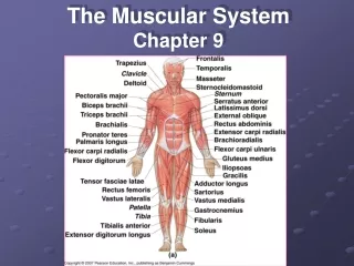
Facial Head Epicranius, frontal belly Temporalis Orbicularis oculi Masseter Zygomaticus Shoulder Orbicularis oris Trapezius Neck Deltoid Sternohyoid Arm Platysma Triceps brachii Sternocleidomastoid Biceps brachii Thorax Brachialis Pectoralis minor Forearm Serratus anterior Pronator teres Pectoralis major Brachioradialis Intercostals Flexor carpi radialis Abdomen Palmaris longus Rectus abdominis Pelvis/thigh Internal oblique Iliopsoas Transversus abdominis Pectineus External oblique Thigh Thigh Rectus femoris Tensor fasciae latae Vastus lateralis Sartorius Vastus medialis Adductor longus Leg Gracilis Fibularis longus Leg Extensor digitorum longus Gastrocnemius Tibialis anterior Soleus Figure 10.4
Neck Epicranius, occipital belly Sternocleidomastoid Arm Trapezius Triceps brachii Shoulder Brachialis Deltoid Forearm Infraspinatus Brachioradialis Teres major Extensor carpi radialis longus Rhomboid major Latissimus dorsi Flexor carpi ulnaris Hip Extensor carpi ulnaris Gluteus medius Gluteus maximus Extensor digitorum Iliotibial tract Thigh Adductor magnus Hamstrings: Leg Biceps femoris Gastrocnemius Semitendinosus Semimembranosus Soleus Fibularis longus Calcaneal (Achilles) tendon Figure 10.5
Muscles of the Head • Two groups • Muscles of facial expression • Muscles of mastication and tongue movement
Muscles of Facial Expression • Insert into the skin • Important in nonverbal communication • All innervated by cranial nerve VII (facial nerve)
Muscles of Facial Expression • Epicranius (occipitofrontalis) • Bipartite muscle consisting of the • Frontalis • Occipitalis • Galea aponeurotica—cranial aponeurosis connecting above muscles • The two muscles have alternate actions of pulling the scalp forward and backward
Epicranius Galea aponeurotica Corrugator supercilii Frontal belly Orbicularis oculi Occipital belly Levator labii superioris Zygomaticus minor and major Temporalis Buccinator Masseter Risorius Sternocleidomastoid Orbicularis oris Trapezius Mentalis Splenius capitis Depressor labii inferioris Depressor anguli oris Platysma Figure 10.6
Muscles of Mastication and Tongue Movement • Four pairs involved in mastication • Prime movers of jaw closure • Temporalis and masseter • Grinding movements • Medial and lateral pterygoids
Muscles of Mastication and Tongue Movement • All are innervated by cranial nerve V (trigeminal nerve) • Buccinator muscles (of facial expression group) also help by holding food between the teeth • Three muscles anchor and move the tongue • All are innervated by cranial nerve XII (hypoglossal nerve)
Temporalis Orbicularis oris Masseter Buccinator (a) Figure 10.7a
Muscles of Mastication and Tongue Movement PLAY A&P Flix™: Temporalis PLAY A&P Flix™: Masseter PLAY A&P Flix™: Buccinator
Lateral pterygoid Medial pterygoid Masseter pulled away (b) Figure 10.7b
Tongue Styloid process Styloglossus Genioglossus Hyoglossus Stylohyoid Mandibular symphysis Hyoid bone Geniohyoid Thyrohyoid Thyroid cartilage (c) Figure 10.7c
Muscles of the Anterior Neck and Throat • Most are involved in swallowing • Two groups • Suprahyoid • Infrahyoid
Suprahyoid Muscles of the Anterior Neck and Throat • Four deep muscles are involved in swallowing (they move the hyoid bone and larynx) • Form the floor of the oral cavity • Anchor the tongue • Move the hyoid bone and the larynx
Infrahyoid Muscles of the Anterior Neck and Throat • Straplike muscles that depress the hyoid and larynx as swallowing ends and during speaking
Median raphe Anterior belly Mylohyoid Digastric Stylohyoid Posterior belly Hyoid bone Omohyoid (superior belly) Stylohyoid (cut) Thyrohyoid Sternohyoid Thyroid cartilage of the larynx Sternocleido- mastoid Thyroid gland Omohyoid (inferior belly) Sternothyroid (a) Figure 10.8a
Tensor veli palatini Levator veli palatini Styloid process Buccinator Superior pharyngeal constrictor Mandible Middle pharyngeal constrictor Mylohyoid (cut) Hyoid bone Geniohyoid Thyrohyoid membrane Hyoglossus Inferior pharyngeal constrictor Thyroid cartilage of larynx (c) Esophagus Trachea Figure 10.8c
Infrahyoid Muscles of the Anterior Neck and Throat PLAY Animation: Rotatable head PLAY Animation: Rotatable face
Muscles of the Neck and Vertebral Column • Two functional groups • Muscles that move the head • Muscles that extend the trunk and maintain posture
Muscles of the Neck and Vertebral Column: Head Movement • Sternocleidomastoid—major head flexor • Suprahyoid and infrahyoid—synergists to head flexion • Sternocleidomastoid and scalenes—lateral head movements • Semispinalis capitis—synergist with sternocleidomastoid • Splenius (capitis and cervicis portions): head extension, rotation, and lateral bending
Base of occipital bone 1st cervical vertebra Mastoid process Middle scalene Sternocleido- mastoid Anterior scalene Posterior scalene (a) Anterior Figure 10.9a
Mastoid process Splenius capitis Spinous processes of the vertebrae Splenius cervicis (b) Posterior Figure 10.9b
Muscles of the Neck and Vertebral Column: Head Movement PLAY A&P Flix™: Splenius capitis PLAY A&P Flix™: Semispinalis capitis
Muscles of the Neck and Vertebral Column: Trunk Extension • Deep (intrinsic) back muscles • Erector spinae (sacrospinalis) group—prime movers of back extension and lateral bending • Iliocostalis • Longissimus • Spinalis • Semispinalis and quadratus lumborum—synergists in extension and rotation
Ligamentum nuchae Mastoid process of temporal bone Semispinalis capitis Longissimus capitis Iliocostalis cervicis Semispinalis cervicis Longissimus cervicis Semispinalis thoracis Iliocostalis thoracis Longissimus thoracis Spinalis thoracis Iliocostalis Erector spinae Longissimus Spinalis Multifidus Iliocostalis lumborum Quadratus lumborum External oblique (d) Figure 10.9d
Muscles of the Neck and Vertebral Column: Trunk Extension PLAY A&P Flix™: Iliocostalis PLAY A&P Flix™: Longissimus PLAY A&P Flix™: Spinalis