Download

1 / 21
430 likes | 1.94k Vues
Vascular Grafts, Stents, and Meshes. Introduction. Arterial diseases Major medical problem world-wide One of the main causes of death in the US Surgical reconstruction Does not deal with the causes of disease Not fully understood Solve problems caused by symptoms. Porosity

E N D
Introduction • Arterial diseases • Major medical problem world-wide • One of the main causes of death in the US • Surgical reconstruction • Does not deal with the causes of disease • Not fully understood • Solve problems caused by symptoms
Porosity Essential component Long-term patency Permit ingrowth of cells (fibroblasts) Necessary for uniform and satisfactory bonding of the internal lining Compliance Must be matched to the properties of the artery Occlusion of the replacement High shear stress at suture line Turbulence of blood flow with local stagnation Biodegradability Control hemorrhage Low porosity during implantation/high porosity during healing Enables patient’s cells to replace the graft with natural tissue Characteristics of Vascular Grafts
Dilitation Permanent enlargement of graft diameter due to pulsing stresses Most frequent mechanical failure 35 months after implantation Suture failure Mismatch of compliances Suture material failure 30-50 months after implantation Defects in the graft Rare Holes, perforations, rents, slits Occur during manufacturing and handling Usually hard to detect Bleeding and infection Rare Usually within first 10 months Suture line Interstices of the graft Graft Failure
1906 to 1916 First documented case of veins used to replace human arteries 1940s Arterial grafts from young, dying persons used (allograft/homograft) Degenerative changes Rejection Abandoned in the 1950s 1950’s Carotid artery from cow (heterograft/xenograft) was initially successful Development of complications 1976 Tanned umbilical vein graft Still used for lower extremity revascularization History – Natural Materials
History - Synthetics • During WW I (1914-1918) paraffin-coated Ag tubes introduced • Also, paraffin-coated glass, aluminum, polyethylene, and steel mesh • Inert • Non-porous • Compliance
Cardiovascular System • Blood vascular system • Distributes nutritive materials, oxygen, and hormones • Removes cellular waste products of metabolism and carbon dioxide • 60,000 miles (http://www.cardio.bayer.com/en/heart_vascular/vascular/)
Heart • Modified blood vessel serving as a double pump • 2 sides, left and right heart • Atria and ventricles • Atria – reservoirs • Ventricles - pumps
Arteries, Capillaries, and Veins • Arteries • Carry blood from the heart to the extremities • Pulsating pressure • Capillaries • Major location of biological interchange • Meshwork of fine tubules • Veins • Return blood from extremities to heart • Constant pressure
Muscular Arteries • Parts of the body under varying conditions require different amounts of blood • Supply arteries must be able to vary the size of their lumina • Walls consist of smooth muscle fibers • Controlled by autonomic nerve system
Arterioles • Blood delivered to capillaries under reduced pressure because walls thin to allow nutrient/waste transfer • Narrow arteries (<100 μm or less) with thick, muscular walls
Microscopic Structure of Arteries • Single layer of endothelial cells • Capillaries – major wall component • Structure and thickness of other walls depends upon function
“Elastic Arteries” • Wall thickness is relatively thin for the size of the vessel • Large arteries
Blood • Belongs to group of tissues called connective tissue • 7% of total body weight • 5 ℓ in average adult • Formed elements (55%) • Red cells, white cells, platelets • Plasma (45%) • Imparts fluid properties to blood
Plasma • Fluid that transports nutritive materials Component Percentage Water 91-92 Protein (fibrinogens, globulins, albumins) 7-8 Other solutes: Small electrolytes (Na+, K+ , Ca2+, Mg2+, Cl-, HCO3 -, PO4 3-, SO4 2-) Nonprotein nitrogen substances [NPN] (urea, uric acid, creatine, creatinine, ammonium salts) Nutrients (glucose, lipids, amino acids) Blood gasses (oxygen, carbon dioxide, nitrogen) Regulatory substances (hormones, enzymes) 1-2
Blood Component Number or percentage Red blood cells (erythrocytes) 4-5x10⁶/mm3 White blood cells (leukocytes) 6000-9000/mm3 Agranular leukocytes: Lymphocytes 30-35% (of leukocytes) Monocytes 3-7% (of leukocytes) Granular leukocytes: Neutrophils 55-60% (of leukocytes) Eosinophils 2-5% (of leukocytes) Basophils 0-1% (of leukocytes) Platelets (thrombocytes) 2-4x10⁵/mm3
Discontinuity in endothelial lining Leads to deposition of proteins Leads to platelet aggregation Followed by adhesion of other platelets Coagulation initiated by factors in plasma Cascade of at least 13 plasma proteins Last step is conversion of monomer fibrinogen to fibrin through action of plasma enzyme thrombin creating a fibrous network that traps blood cells Blood Clotting
Angioplasty • Opening up plaque-narrowed artery without doing major surgery • Catheter w/balloon tip inserted into coronary or major leg or arm artery • Inflation of balloon • often repeatedly • stretches artery wall • disrupts plaques
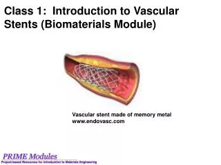
Stents • wire mesh • Used in 70-90% of all angioplasty procedures • keeps the vessel open after widening • placed onto balloon prior to insertion • permanently attached as balloon inflates
Open narrowed or blocked coronary artery in patients suffering from angina – alternative to bypass surgery Open a blocked artery in the pelvis, leg, or arm – peripheral arterial disease Control blood pressure in renal hypertension – caused by narrowing of one or both arteries supplying kidneys To keep blood vessel grafts open in patients undergoing hemodialysis – most have a graft between a artery and vein in the arm to easily draw and replace blood maintain blood flow to the brain by keeping open the carotid artery Uses of the Procedure