Download
1 / 48
490 likes | 750 Vues
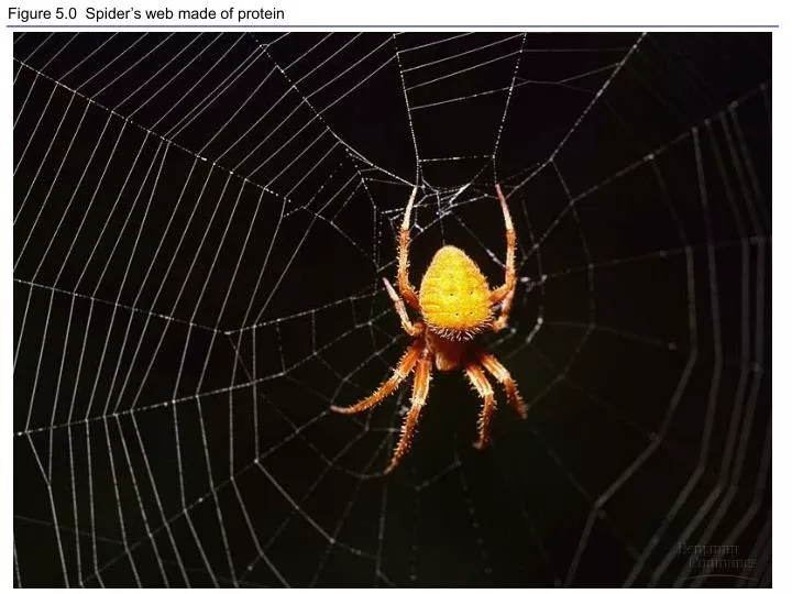
Figure 5.0 Spider’s web made of protein. Figure 5.1 Building models to study the structure and function of macromolecules. Figure 5.2 The synthesis and breakdown of polymers. Figure 5.3 The structure and classification of some monosaccharides.

E N D
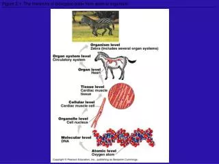
Figure 5.1 Building models to study the structure and function of macromolecules
Figure 5.3 The structure and classification of some monosaccharides
Figure 5.29 The components of nucleic acids; differences between DNA and RNA
Figure 5.3x Hexose sugars Glucose Galactose
Figure 5.5x Glucose monomer and disaccharides Glucose monomer Sucrose Maltose
Figure 5.7x Starch and cellulose molecular models Glucose Glucose Cellulose Starch
Figure 5.9 Chitin, a structural polysaccharide: exoskeleton and surgical thread
Figure 5.10 The synthesis and structure of a fat, or triacylglycerol
Figure 5.11x Saturated and unsaturated fats and fatty acids: butter and oil
Figure 5.11 Examples of saturated and unsaturated fats and fatty acids
Figure 5.13 Two structures formed by self-assembly of phospholipids in aqueous environments
Figure 8.6 The detailed structure of an animal cell’s plasma membrane, in cross section
Figure 4.8 A comparison of functional groups of female (estradiol) and male (testosterone) sex hormones
Figure 5.15 The 20 amino acids of proteins: polar and electrically charged
Figure 5.22 Examples of interactions contributing to the tertiary structure of a protein
Figure 5.19 A single amino acid substitution in a protein causes sickle-cell disease
LE 5-21b Sickle-cell hemoglobin Normal hemoglobin Primary structure Primary structure Val Val His His Thr Pro Glu Glu Thr Pro Val Glu Leu Leu 1 1 2 4 6 2 4 6 7 7 3 5 3 5 Exposed hydrophobic region Secondary and tertiary structures Secondary and tertiary structures b subunit b subunit a a Quaternary structure Sickle-cell hemoglobin Normal hemoglobin (top view) Quaternary structure a a Function Molecules do not associate with one another; each carries oxygen. Molecules interact with one another to crystallize into a fiber; capacity to carry oxygen is greatly reduced. Function
Figure 5.28 DNA RNA protein: a diagrammatic overview of information flow in a cell
Figure 5.29 The components of nucleic acids; differences between DNA and RNA
Table 5.2 Polypeptide Sequence as Evidence for Evolutionary Relationships