Major Histocompatibility Complex (MHC)
Major Histocompatibility Complex (MHC). Department of Microbiology. Major Histo -compatibility Complex (MHC). Major Histocompatibility complex (MHC) is a cluster of tightly linked genes (crossing over frequency is very low i.e., 0.5%)

Major Histocompatibility Complex (MHC)
E N D
Presentation Transcript
Major Histocompatibility Complex (MHC) Department of Microbiology
Major Histo-compatibility Complex (MHC) • Major Histocompatibility complex (MHC) is a cluster of tightly linked genes (crossing over frequency is very low i.e., 0.5%) • They code for protein molecules playing role in presentation of processed antigen fragments and in discrimination between self and non-self. • Two main products encoded by MHC genes are MHC I and MHC II molecules present on the cell surface and playing role in presentation of antigen fragments to immune cells. • Cluster of genes found in all mammals • Its products play role in discriminating self/non-self • Participant in both humoral and cell-mediated immunity
MHC I Molecule • In general, MHC I molecules are present on all nucleated cells. • MHC I receptors are made up of two polypeptide chains called alpha chain and beta-macroglobulin. • α- chain is of 45 kDa whereas β-macroglobulin is of 12 kDa in size. • The alpha chain is encoded by genes of MHC I where as β- macroglobulin is coded by genes other than MHC.
MHC I Molecule • The alpha chain is made up of 3 extracellular domains i.e., α 1, α 2 and α 3 domains. • The peptide binding clefts are formed by two distal domains of α chain i.e., α1 and α 2 domains. • This peptide binding cleft is close ended and can bind to a peptide of 8-9 amino acids.
MHC I Molecule • The antigen fragments loaded on MHC I molecules are derived from processing of endogenous antigens. • Endogenous antigens are antigens that are synthesized within the cell itself . • Examples of Endogenous antigens are normal self antigens, altered self antigens (tumour antigens), viral antigens etc. • Note that the viruses utilize the host cell machinery to produce viral proteins. Thus, viral proteins are also considered as endogenous antigen.
MHC I Molecule • The endogenous antigens are processed through “cytosolic pathway” of antigen processing. • The antigenic peptides loaded on MHC I molecules are presented to T cells having CD8+ receptors. • (CD 8+ positive cells are T cytotoxic cells). Cytosolic pathway of antigen processing
MHC II molecule • In general, MHC II molecules are present especially on professional antigen presenting cells (APCs) i.e., Macrophages, Dendritic cells and B cells. • MHC II receptors are made up of two polypeptide chains called alpha and beta-chains. • α-chain is of 31 -34 kDa in size. • β-chain is of 25-29 kDa in size.
MHC II molecule • The alpha chain is made up of 2 extracellular domains i.e., α 1, and α 2 domains. • The beta chain is made up of 2 extracellular domains i.e., β 1, and β 2 domains. • The peptide binding clefts are formed by two distal domains of α and β chains i.e., α1 and β1 domains. • This peptide binding cleft is open ended and can bind to a peptide of 18-20 amino acids in size.
MHC II molecule • The antigen fragments loaded on MHC II molecules are derived from processing of exogenous antigens. • Exogenous antigens are engulfed by the cells from outside and they are taken into vacuoles. • The exogenous term is used with respected to the APCs which performs the processing and presentation of antigen. • The term generally includes bacterial antigens, parasitic antigens and proteins released from even other self cells.
MHC II molecule • The exogenous antigens are processed through “endocytic pathway” of antigen processing. • The antigenic peptides loaded on MHC II molecules are presented to T cells having CD4+ receptors. • (CD 4+ positive cells are T helper cells). Endocytic pathway of antigen presentation
Major Histocompatibility complex in different Species • All vertebrates have MHC (Gene clusters) • MHC of different species are named as shown in table.
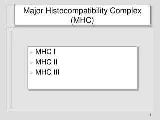
Organization of MHC gene • Genes of MHC are organized in 3 groups: • Class I MHC genes • Encode for glycoprotein molecules expressed on the surface of all nucleated cells • Major function to present processed Antigens to TC cells. • Class II MHC genes • Encode for glycoprotein molecules expressed on M, B-cells, DCs (mainly on professional APCs) • Major function to present processed Antigens to TH cells • Class III MHC genes • Encode for secreted proteins that have immune functions. Ex. Complement proteins, inflammatory molecules • Do not play role in presentation of antigenic peptides
Organization of MHC gene in Human • Class I MHC Genes Found In Regions A, B and C (codes for α-chain of MHC I molecule). (It is important to note that β-macroglobulin component of MHC I is not coded by MHC genes) • Class II MHC Genes Found In Regions DR, DP and DQ (codes for both α and β chains of MHC II molecule). • Class III MHC codes for a few complement components (C2, C4, factor B) and cytokines for eg., TNF-α and β
MHC genes are polymorphic • MHC Products Are Highly Polymorphic • Vary considerably from person to person • Crossing over frequency (0.5%) between MHC genes is very less. • Thus, MHC alleles present on one chromosome tends to remain as such and inherited as set. • This set of MHC alleles is generally termed as “Haplotype”. • Thus, a person have two haplotypes transferred from mother and father. • MHC Alleles Are Co-dominantly Expressed • Both mother and father alleles are expressed.
MHC-peptide binding • Binding of processed peptide with the MHC does not show high level of specificity (as shown in antigen-antibody interaction or antigen-T cell receptor interactions). • Peptide of suitable size with certain amino acid residues at specific positions may bind to the MHC molecule. • For eg: Peptides that bind to MHC molecule I is found to be of 8-10 amino acid. • And all these peptides have specific amino acid residues that is essential for binding to a particular MHC molecule. • Such binding specificity exhibited by MHC molecules for peptides is known as “promiscus” binding. • It is estimated that one type of MHC molecule (molecules from one allele) can bind to 2500 different types of peptide.
All these peptides of 9 aa in length can bind to H-2Dd (MHC I molecule of mice). In this example binding specificity is decided by amino acid present at position 2, 3 and 9. So any peptide of this size with Glycine, Proline at 2nd and 3rd position and Leucine or Isoleucine at 9th position can bind to this MHC molecule.
MHC haplotypes and disease resistance • MHC plays very important role in giving protection by presenting epitopes against which immune response is protective. • Thus, MHC alleles are important factor which determines susceptibility or resistance against any infectious agent. • For this reason MHC exhibit high level of polymorphism and each allele binds to a different set of polypeptides. • In cattle, BoLA-A*6 allele is associated with resistance to mastitis. • BoLA-DRB3.2*23 allele is associated with incidence of severe coliform mastitis. • Poultry: Resistance against Mareks disease is associated with _______________________________________.
Class I And II Specificity • Several Hundred Allelic Variants Have Been Identified In Humans • However, up to 6 MHC I And 12 MHC II Molecules Are Expressed In An Individual • Enormous Number Of Peptides Needs To Be Presented Using These MHC Molecules • To Achieve This Task MHC Molecules Are Not Very Specific For Peptides (Unlike TCR and BCR) • Promiscuous Binding Occurs • A peptide can bind a number of MHC • An MHC molecule can bind numerous peptides
Class I And II Diversity And Polymorphism • MHC Is One Of The Most Polymorphic Complexes Known • Alleles Can Differ Up To 20 a/a • Class I Alleles In Humans: 240 A, 470 B, 110 C • Class II Alleles In Humans: HLA-DR 350 , 2 ! • HLA-DR • genes vary from 2-9 in different individuals!!!, • 1 gene ( can combine with all products increasing number of APC molecules) • DP (2 , 2 ) and DQ (2 , 3 )
Alphachain Beta chain Variable region “V” CHO CHO CHO CHO Constant region “C” Hinge “H” Disulfide bridge + Transmembrane region + + Cytoplasmic tail Structure of T Cell Receptor
α1 NH2 NH2 Alloantigenic sites β2 NH2 α2 COOH CHO α3 Disulfide bridge Papain cleavage Plasma membrane OH P Cytoplasm COOH Structure of Class I MHC
NH2 NH2 α1 β1 CHO CHO α2 β2 CHO Plasma membrane Cytoplasm COOH COOH Structure of Class II MHC
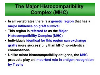
Histocompatibility (transplantation) antigensAntigens on tissues and cells that determine their rejection when grafted between two genetically different individuals • Major histocompatibility (MHC) antigensHistocompatibility antigens that cause a very strong immune response and are most important in rejection • MHC complexGroup of genes on a single chromosome encoding the MHC antigens • HLA (human leukocyte antigens)MHC antigens of man (first detected on leukocytes) • H-2 antigensMHC antigens of mouse • Types of graft (figure 1) • XenograftGrafts between members of different species (also known as heterologous, xenogeneic or heterografts) • AllograftGrafts between two members of the same species (also known as allogeneic or homograft) • IsograftGrafts between members of the same species with identical genetic makeup (identical twins or inbred animals) • HaplotypeA group of genes on a single chromosome
PRINCIPLES OF TRANSPLANTATION (figure 2) • An immunocompetent host recognizes the foreign antigens on grafted tissues (or cells) and mounts an immune response which results in rejection. On the other hand, if an immunocompromised host is grafted with foreign immunocompetent lymphoid cells, the immunoreactive T-cells in the graft recognize the foreign antigens on the host tissue, leading to damage of the host tissue. • Host-versus-graft-reactionThe duration of graft survival follows the order, xeno- < allo- < iso- = auto- graft. The time of rejection also depends on the antigenic disparity between the donors and recipient. MHC antigens are the major contributors in rejection, but the minor histocompatibility antigens also play a role. Rejection due to disparity in several minor histocompatibility antigens may be as quick or quicker than rejection mediated by an MHC antigen. As in other immune responses, there is immunological memory and secondary response in graft rejection. Thus, once a graft is rejected by a recipient, a second graft from the same donor, or a donor with the same histocompatibility antigens, will be rejected in a much shorter time. • Figure 3 Graft versus host disease Graft-versus-host (GVH) Reaction • Histocompatible lymphoid cells, when injected into an immunocompromised host, are readily accepted. However, the immunocompetent T lymphocytes among the grafted cells recognize the alloantigens and, in response, they proliferate and progressively cause damage to the host tissues and cells. This condition is known as graft-versus-host (GVH) disease (figure 3) and is often fatal. • Common manifestations (figure 4) of GVH reaction are diarrhea, erythema, weight loss, malaise, fever, joint pains, etc. and ultimately death.