Download
1 / 38
710 likes | 2k Vues
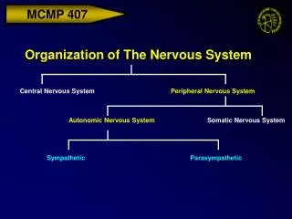
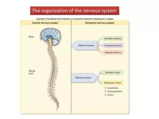
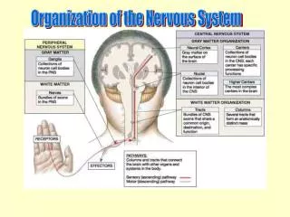
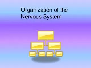
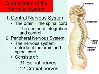
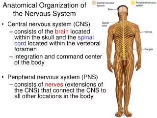
Central nervous system (CNS) consists of the brain located within the skull and the spinal cord located within the vertebral foramen integration and command center of the body Peripheral nervous system (PNS)

E N D
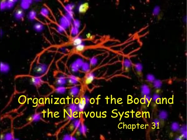
Central nervous system (CNS) consists of the brain located within the skull and the spinal cord located within the vertebral foramen integration and command center of the body Peripheral nervous system (PNS) consists of nerves (extensions of the CNS) that connect the CNS to all other locations in the body Anatomical Organization of the Nervous System
Nervous System • One of 2 controlling and communicating systems of the body (other is the endocrine system) • Transmit sensory information • propagate APs to the CNS following a stimulus which has changed a variable from its set point • from eyes, skin, blood vessels, ears, digestive tract, joints, muscles, lungs… • Integration • interpretation of sensory information by the CNS • type, location and magnitude of stimulus • Transmit motor information • propagate APs from the CNS to various effector organs throughout the body • provides a way to respond to stimuli
Cells of the Nervous System • The two principal cell types of the nervous system are: • Neurons • hundreds of thousands of neurons extend axons and make synapses all over the body with other neurons, muscles and glands • communicate through action potentials • allows for short response times to changes in homeostasis • Neuroglia • guide developing neurons to make synapses • provide a supportive scaffolding for developed neurons
Neuron Types of the Nervous System • Sensory (afferent) • associated with sensory receptors • propagate APs via the PNS toward the CNS • Interneurons • integrate information within the CNS • receive APs from sensory neurons and initiate APs in motor neurons • Motor (efferent) • propagate APs via the PNS away from the CNS • All 3 neuron types are used to respond to stimuli • reflex
Some neurons in the CNS are myelinated, while most are unmyelinated All of the neurons in the PNS are myelinated Areas of the CNS that are made of myelinated neurons are called white matter represent the locations of long sensory and motor neurons Areas of the CNS that are made of unmyelinated neurons are called gray matter represent the locations of short interneurons which make many synapses for integration to process sensory information and initiate motor information Myelination of Neurons of the Nervous System
Spinal Cord The spinal cord is attached to the brain and extends to the lumbar region of the vertebral column Functions include: integration of basic stimuli presented to the body below the neck through simple reflexes withdrawal reflex in response to pain sending sensory and motor information to and from the brain
Spinal Cord Anatomy Dorsal (posterior) horns (left and right) sensory information enter the cord on the dorsal aspect where they synapse with interneurons or motor neurons extend into dorsal roots and ganglia(group of cell bodies outside the CNS) Ventral (anterior) horns (left and right) motor information exits the cord on the ventral aspect where they control effectors (muscle or glands) extend into motor roots Dorsal and ventral roots merge together to form spinal nerves
Cerebral Cortex 4 lobes frontal, parietal, temporal and occipital location of interneurons for perception of all senses site of memory, emotion, learning site of initiation of voluntary skeletal muscle contraction
The Cerebellum Protrudes under the occipital lobes of the cerebrum Makes up 11% of the brain’s mass Modifies the motor information leaving the motor cortex provides precise timing and appropriate patterns of skeletal muscle contraction to maintain balance and coordination Cerebellar activity occurs subconsciously
Brain Stem Comprised of the pons and the medulla oblongata Clusters of neurons (brain centers) in regions of the pons and medulla control the basic life functions: heart rate controlled by the cardioacceleratory and cardioinhibitory centers in the medulla blood pressure controlled by the cardioacceleratory, cardioinhibitory, and vasomotor centers in the medulla breathing rate controlled by the inspiratory and expiratory centers in the medulla and pons, respectively Control of effectors occurs through the Autonomic Nervous System
Peripheral Nervous System The PNS consists of nerves (bundles of axons) propagate APs to and away from the CNS 12 pairs (left and right) of cranial are connected to the brain and 31 pairs (left and right) of nerves are connected to the spinal cord Sensory (afferent) all axons carry impulses from sensory receptors via the PNS to the CNS Motor (efferent) all axons carry impulses via the PNS from CNS Mixed a mixture of sensory and motor neurons that carry impulses via the PNS to and from CNS most common type of nerve in the body
Nerves Nerve cordlike organ of the PNS consisting of axons enclosed by connective tissue Connective tissue coverings include: Endoneurium loose connective tissue that surrounds each individual axon Perineurium coarse connective tissue that bundles axons into fascicles Epineurium tough fibrous connective tissue around a nerve
Reflexes • A rapid, predictable motor response to a stimulus • Reflexes can be: • simple • involve peripheral nerves and the spinal cord • rapid • learned (acquired) • involve peripheral nerves and require thought • slower • Following a stimulus, the sensory and motor information of a reflex follows a pathway called a reflex arc • in many spinal reflexes, the effector is nearby the location of the stimulus
Reflex Arc • There are five components of a reflex arc • Receptor • detect stimulus • Sensory neuron • transmits the afferent impulse to the CNS • Integration (control) center • region within the CNS where synapses (processing of sensory info) occur • Motor neuron • sends efferent information to an effector • Effector • muscle fiber or gland that responds to the efferent impulse • the activity of the effector depends upon the magnitude of the stimulus
Sensory Receptors • Structures specialized to respond to stimuli: • nerve endings (dendrites of neurons) • sense organs • nerve endings combined with other tissue types to enhance detection of a stimuli • example: taste buds • Mechanoreceptors • respond to touch, pressure, stretch and itch • Thermoreceptors • respond to changes in temperature • Photoreceptors • respond to light • Chemoreceptors • respond to chemicals • Nociceptors • respond to pain
Neural Integration of the CNS • Qualitative information (salty, pain or temperature) depends upon which neurons are propagating APs • Quantitative (strength) information depend on: • the number of neurons that are firing APs • the frequency of APs fired per neuron
Sensory Division of the Peripheral NS Sensory division • made of afferent neurons • somatic • sensory neurons send APs from skin, skeletal muscles, and joints • visceral • sensory neurons send APs from organs within the abdominal and thoracic cavaties
Motor Division of the Peripheral NS Motor division • made ofefferent neurons control the action of effectors • somatic • motor neurons send APs to voluntaryskeletal muscle • visceral • motor neurons send APs to involuntarycardiac muscle, smooth muscle and glands • a.k.a. the Autonomic Nervous System (ANS) • 2 antagonistic (opposing) divisions • Sympathetic • Parasympathetic • the two divisions control the same effectors (with few exceptions) but create opposite responses in the effectors
Motor Pathways of the Somatic Nervous Division vs. Autonomic Nervous Division
Autonomic Nervous System • Visceral motor neurons of the Peripheral NS control the activity of involuntary effectors such as cardiac muscle, smooth muscle and glandular secretion affecting: • heart rate • breathing rate • sweating • digestion • blood pressure • Action potentials in these motor neurons are initiated in the medulla oblongata and the pons • these motor neurons exit the brain by: • descending tracts of the spinal cord • exit spinal cord via spinal nerves • cranial nerves
Function of the Sympathetic Division • The sympathetic division is called the “fight or flight” system • activated when the body needs to expend energy • Involves E activities • exercise, excitement, emergency, and embarrassment • Promotes necessary changes during these activities • increases heart rate, blood pressure, respiration rate, blood flow to skeletal muscles, glucose metabolism • decreases the activity of and blood flow to the digestive system organs • Its activity is illustrated by a person who is threatened
Function of the Parasympathetic Division • The parasympathetic nervous system is called the “rest and digest” system • activated when the body needs to conserve energy • Involves the Dactivities • digestion, defecation, and diuresis (urination) • Promotes necessary changes during these activities • decreases heart rate, blood pressure, respiration rate, blood flow to skeletal muscles, glucose metabolism • increases the activity of and blood flow to the digestive system organs • Its activity is illustrated in a person who relaxes after eating a meal
Efferent Pathways of the ANS • Efferent pathways of the ANS consist of a two-neuron chain between the brain or spinal cord and the effector • synapses between the neurons occur at ganglions • The cell body and dendrites of the preganglionic neuron is located in the CNS and the axon extends along a nerve to the ganglion • The cell body and dendrites of the postganglionic neuron is located in the ganglion and the axon extends to an effector organ
Motor Pathways of the Somatic Nervous Division vs. Autonomic Nervous Division • All somatic motor neurons exocytose ACh • ACh binds to nicotinic acetylcholine receptors on the skeletal muscle fiber leading to its contraction • All preganglionic motor neurons exocytose ACh • ACh binds to nicotinic acetylcholine receptors on the postganglionic neuron creating an AP • All parasympathetic postganglionic motor neurons exocytose ACh • ACh binds to muscarinic acetylcholine receptors on the effector tissue/organ causing a response • All sympathetic postganglionic motor neurons exocytose norepinephrineNE • NE binds to adrenergic receptors on the effector tissue/organ causing a response
Effects of Neurotransmitters of the ANS • The way the 2 divisions of the ANS can create opposite responses in the effectors that they control is by the release of different neurotransmitters onto the cells of the effectors • The cells of each organ controlled by the ANS have membrane receptors to BOTH ACh and NE • organs are dually controlled • The response of the organ is determined by the identity of the neurotransmitter released • the binding of ACh to its receptor will cause the effector to respond in one way • the binding of NE to its receptor will cause the effector to respond in the opposite way • The effect of ACh and NE is effector specific • NE increases heart rate, ACh decreases heart rate • NE decreases the secretion of saliva, ACh increases the secretion of saliva