DVT-PE
DVT-PE. Nitin Bhatt MD Director, ICU VAMC, Reno.

DVT-PE
E N D
Presentation Transcript
DVT-PE Nitin Bhatt MD Director, ICU VAMC, Reno
Q;3, A 45-year-old woman is evaluated in the emergency department because of the sudden onset of shortness of breath and chest tightness earlier this afternoon. She has been in excellent health and returned 2 days ago from a trip to Japan. On physical examination, her pulse rate is 116/min and respiration rate is 36/min. Cardiopulmonary examination is otherwise unremarkable, and a chest radiograph shows no infiltrates. Measurement of arterial blood gases on room air shows the following: pH 7.48, Paco2 24 mm Hg, and Pao2 78 mm Hg; oxygen saturation is 96%. What is the most appropriate immediate next step in this patient’s management? ( A ) Pulmonary angiogram( B ) Compression ultrasonography of the lower extremities( C ) Therapy with diazepam( D ) Intravenous heparin( E ) Ventilation-perfusion lung scan
DVT-PE • Different stages of same disease • 90%-95% of PE originate from clots in lower extremities • 600,000 per year remain undiagnosed in USA as signs and symptoms are nonspecific
Asymptomatic PE in DVT pts • About 40% of patients with venogram proven DVT had asymptomatic PEs when investigated by pulmonary angiogram. Ken Moser et al
Of an estimated 200,000 deaths per year in the United States, only 13,000 (6%) occur in patients who have received treatment. The vast majority of patients (94%) who die of pulmonary embolism do not receive treatment because the diagnosis is not made
PE Difficulty of Diagnosis • Multiple clinical presentations • Nonspecific signs and symptoms Mortality Rates • With treatment: 5% to 8% (more recent data suggests 2-3%) • Without treatment: 25% to 30%
Q;3, A 45-year-old woman is evaluated in the emergency department because of the sudden onset of shortness of breath and chest tightness earlier this afternoon. She has been in excellent health and returned 2 days ago from a trip to Japan. On physical examination, her pulse rate is 116/min and respiration rate is 36/min. Cardiopulmonary examination is otherwise unremarkable, and a chest radiograph shows no infiltrates. Measurement of arterial blood gases on room air shows the following: pH 7.48, Paco2 24 mm Hg, and Pao2 78 mm Hg; oxygen saturation is 96%. What is the most appropriate immediate next step in this patient’s management? ( A ) Pulmonary angiogram( B ) Compression ultrasonography of the lower extremities( C ) Therapy with diazepam( D ) Intravenous heparin( E ) Ventilation-perfusion lung scan
Q:3, Ans=D, When venous thromboembolism is strongly suspected, the first action taken should be to administer heparin in a therapeutic dose (in the absence of contraindications to anticoagulation) to prevent new clot formation, allow unopposed fibrinolytic activity to facilitate thrombus resolution, and avoid progression into the pulmonary circulation. Therapy should be started before performing confirmatory diagnostic tests such as a ventilation-perfusion scan, pulmonary angiogram or compression ultrasonography of the lower extremities. Subtherapeutic or delayed anticoagulation is associated with unacceptable recurrence rates of venous thromboembolism, and is a common error in the initial phases of treatment. Although relatively less heparin is required to prevent the coagulation cascade from being initiated, more is required after the cascade is under way. Therefore, early administration of an adequate dose is required. For continuous infusions, the activated partial thromboplastin time should be monitored regularly to ensure that the dose is therapeutic. Inadequate initial heparin therapy also has been associated with late recurrences of thromboembolism. Anxiolytic therapy would not be an appropriate as the initial step in this patient’s manageme
Virchow`s Triad • Venous stasis • Injury to the intima • Change in the coagulability of blood
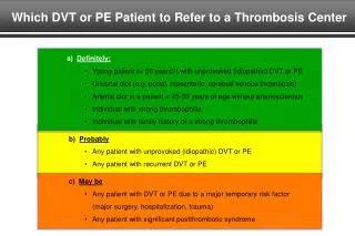
Acquired Risk Factors for VTE • Surgery and Trauma • Prolonged Immobilization • Age greater than 40 • Obesity, Inflammatory Bowel Disease, CHF • Cancer, PNH, Nephrotic syndrome • Myeloproliferative Disorder • Previous DVT or PE, Air-travel • Pregnancy and puerperium • Contraceptives or Hormone Replacement Therapy • Resistance to Activated Protein-c (not F-5 Leiden) • Lupus Anticoagulant/ Antiphospholipid antibody • Mild-moderate Hyperhomocystinemia
Factor V Leiden and VTE • 9253 randomly selected Danish people, heterozygous and homozygous F V Leiden was associated with hazard ratio for VTE of 3 and 18 respectively, compared to non-carrier population. • Simultaneous presence of smoking, obesity and old age increased 10 year absolute risk of VTE to 10% and 51% in Heterozygous and homozygous, resp. Juul et al, AIM 2004; 140:330-337
Natural history • DVT originates as a platelet nidus near venous valves of lower exremities and grows by accretion of platelets and fibrin • Newly formed clot more likely to get detached to cause embolism to the FILTER organ, lung, or may be lysed by endogenous lytics • Organization of venous clots cause damage to venous valves and lead to post-phlebitic syndrome
P.E.:Physiology • Hypoxemia in majority, leading to respiratory alkalosis • V/Q mismatch is predominant cause of hypoxemia, some RL shunting occurs • Serotonin from platelets PVR • Bronchoconstriction due to mediators from thrombii
P.E.: physiology • If no pre-existing cardio-pulmo disease, obstruction of 25-30% of pulmo vascular bed leads to PHTN, >75% obstruction will lead to acute RV failure as normal RV can not generate pressure >50 mm Hg • Lack of increase in CVP with large P.E. may suggest severe RV failure
PHTN after first episode of PE • Excluding patients with persistent risk factors for VTE, in a prospective study of idiopathic or transient risk factor associated PE, about1% risk of chronic thromboembolic pulmonary hypertension was noted, confirmed by pulmonary angiography, 14 to 22 months after acute PE- Becattini et al, Chest 2006;130: 172-5
Q-9:52 yr woman, 1 yr h/o dyspnea. Heavy smoker in past, h/o HTN.O/E loud P2, elevated JVP,mild bilat ankle edema. CBC, Chem-panel, HIV, ANA , Rh. Titer negative. CXR: prominent Pulm. Arteries, echo: normal EF, RVE, PA-syst pressure 60, PFTs: normal volumes, DLCO 40% pred, V/Q: mild heterogeneity of perfusion, Swan Ganz: RAP 10, RVP 50/10, PAP 50/20, PAOP 26, CI 2. ?DX 1, LV diastolic dysfunction 2, Chronic Pulmonary embolism 3, Primary pulmonary HTN 4, Pulmonary veno-occlusive disease 5, Constrictive pericarditis
DVT: Clinical diagnosis ? • Clinical suspicion based on symptoms e.g. leg edema, calf pain, etc., only 30% of clinically suspected to have DVT, ultimately have diagnosis proven. • Homan`s sign is not specific or sensitive for DVT • Myositis, cellulitis, hematoma and edmatous state are commonly included in differential diagnosis of DVT
Fedullo & Tapson,NEJM9-25-03Low=10%, Inter=30% and High=>70%
DVT • Contrast venography • Compression ultrasound • Impedence plethysmography
Compression ultrasound (Goldhaber)Artery compressed, but vein did not: DVT
CUS in suspected PE? • Retrospective analysis of data from prospective study, showed that in patients proven to have PE, >60% had positive CUS. Age >70 and signs and symptoms of DVT were independent predictors of positive CUS Girard et al, Chest 2005, P 1593-00
MRI in Dx Of DVT • 101 Pts who underwent venogram for diagnosis of dvt were included.All pts with positive venogram and ¼ of those with negative venogram included • Magnetic Resonance Direct Thrombus Imaging (MRDTI) was used in all patients within 48 hrs of venography and interpreted by 2 reviewers • Sensitivities and specificities were >90 %. Even isolated calf thrombii had 82-92 % sensitivities. Fraser et al, Ann Int Med 1-15-02
Pelvic DVT diagnosis by MRA • 24 patients with proven PE and negative CUS, were studied. • 29% had pelvic DVT • Common iliac vein involved in 5 patients. Stern et al, Chest 2002;122: 115-21
SYMPTOMS AND SIGNS IN 117 PATIENTS WITH ACUTE PULMONARY EMBOLISM WITHOUT PRE-EXISTING CARDIAC OR PULMONARY DISEASE (Stein, Chest 1991)
During fibrinolysis of fibrin, plasmin cleaves factor XIIIa–cross-linked fibrin into an array of intermediate forms. The D-dimer and E fragments are the result of terminal fibrin degradation., P.Bockenstedt,NEJM 9-25-03
Plasma D-Dimer • Sensitive but not a specific test for PE, therefore use for excluding PE, not useful to make the diagnosis. • Not recommended as stand alone test, use with either clinical criteria or V/Q scan or Ultrasound, several diagnostic algorhythms now reported using D-dimer
D-Dimer in diagnosis of VTE:AJRCCM, Feb 2002; 345-8 • Strong correlation noted between location of P.E. and D-Dimer concentration, p<0.001 • Sensitivity of D-Dimer was 93% for excluding segmental or larger P.E. but only 50% for subsegmental P.E.
D-Dimer and Ultrasound in DVTWells et al,NEJM 9-25-03 • Suspected PE cases were excluded • All patients had clinical stratification as high or low likelihood for DVT • Low clinical suspicion and negative D-Dimer rules out DVT • High clinical suspicion but negative D-Dimer and negative US of legs rules out DVT • 39% in D-Dimer group did not need ultrasound, but overall prevalence of DVT-PE was only 15.7%
D Dimer and DVT • Low clinical probability and negative D Dimer value rules out DVT effectively. • High or intermediate clinical probability makes D dimer value unhelpful • Moderate clinical probability and negative D dimer by HIGH sensitivity assey can have DVT ruled out. (Meta-analysis by Wells et al JAMA Jan 11 2006 p 199-207)
D dimer in evaluation of PE • Low clinical probability combined with negative D dimer (SimpliRED whole blood agglutination test) had negative predictive value of 99.5% for excluding PE in a prospective cohort study of ER patients. Wells et al AIM 2001;135:98-107 • In a prospective randomized trial ( Kearon et al, AIM; 2006 p 812-21) low clinical probability and negative d-dimer was associated with <.5% incidence of VTE on follow up. 50% of out-pt and 20% of inpatient fell into this category.
D dimer and PE • Negative simplify D-dimer test and a low clinical probability of PE, resulted in 0.7% incidence of VTE on 90 day follow up of ER patients. Kline et al Chest; June 2006, 1417-23
PE CXR • In majority of cases of PE, cxr is abnormal but nondiagnostic e.g. Atelectasis, Small pleural effusion • Enlarged right descending PA, Decreased pulmonary vascularity (Westermark sign) • Wedge-shaped peripheral infiltrate (Hampton`s hump) • Normal
Chest radiograph showing pulmonary infarct in the right lower lobe. This patient had low-grade fever, hemoptysis, and pleuritic chest pain. The ventilation-perfusion scan was read as high probability for pulmonary embolism. A pleural-based density in the lower lobe with the convexity directed toward the hilum signifies pulmonary infarction. This sign is also known as “Hampton’s hump.”From Bone`s Atlas