Facial Nerve
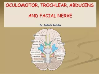
Facial Nerve. Prof. Dr. Norberto V. Martinez Faculty of Medicine and Surgery University of Santo Tomas. Six Anatomical Segments. Intracranial Meatal Labyrinthine Tympanic Mastoid extratemporal. Facial Nerve Surgery & Decompression. 4 functional components. Motor nucleus (efferent)

Facial Nerve
E N D
Presentation Transcript
Facial Nerve Prof. Dr. Norberto V. Martinez Faculty of Medicine and Surgery University of Santo Tomas
Six Anatomical Segments Intracranial Meatal Labyrinthine Tympanic Mastoid extratemporal
4 functional components Motor nucleus (efferent) Parasympathetic fibers-greater superficial petrosal nerve & chorda tympani ( Nervus Intermedius) Special Visceral Afferent from Nucleus Tractus Solitarius(afferent) General Sensory Afferent-cutaneous sensation to external ear & postauricular area (afferent)
Supra nuclear pathway Motor function origin begins at cerebral cortex Primary somatomotor cortex in the precentral gyrus (brodmann area4,6,8)
Facial Nucleus and Brainstem Facial nucleus lies within the reticular formation at the lower level of the pons There is distinctly ipsi & contalateral cortical input within the facial nucleus superior or ventral – receives bilateral input inferior or dorsal – receives contralateral input
INTERNAL AUDITORY CANAL(meatal) Traverse crest divides IAC into superior and inferior Superior portion facial nerve anteriorly superior vestibular nerve posteriorly Inferior portion cochlear nerve anteriorly inferior vestibular nerve posteriorly
FALLOPIAN CANAL Facial canal is approximately 30 mm long From Bills bar up to the stylomastoid foramen 3 intratemporal region labyrinthine tympanic mastoid
Labyrinthine segment Shortest segment (3-4mm) Lies between labyrinth and cochlea Beginning from fundus of IAC extending upto geniculate ganglion* Narrowest portion of fallopian canal is the meatal foramen (junction bet IAC and Labyrinthine segment) Labrynthine segment terminates in the genicultae ganglion and will make a 40 to 80 turn(1st genu)
Mastoid Segment From 2nd genu to stylomastoid foramen Descends inferiorly and becomes more lateral * 2 branches- nerve to stapedius and chorda tympani Angle between chorda tympani and vertical portion is 30 degrees(facial recess)
Extra Temporal Segment 3 minor branches after leaving the stylomastoid foramen post auricular nerve branch to digastric muscle stylohyoid muscle Further arborization occurs with frequent anastomosis occurs in the intraparotid course Five classic branches- temporal,zygomatic,buccal,mandibular,cervical
Blood Supply Blood supply is segmented derived from 3 arterial sources Nager 1953 brainstem to IAC: AICA perigeniculate segment: Mid. meningeal artery mastoid –tympanic: stylomastoid branch of post auricular artery
House Brackmann Facial Nerve Grading System I. Normal • Normal facial function in all areas
House Brackmann Facial Nerve Grading System II. Mild Dysfunction • Gross • Slight weakness noticeable in close inspection . May have very slight synkinesis. At rest normal symmetry and tone. • Motion • Forehead: moderate to good function • Eye: complete closure with minimal effort • Mouth: slight assymetry
House Brackmann Facial Nerve Grading System III. Moderate Dysfunction • Gross • Obvious, but not disfiguring difference between the two sides. Noticeable but not severe synkinesis, contracture, or hemifacial spasm. At rest, normal symmetry and tone. • Motion • Forehead: slight to moderate movement • Eye: complete closure with effort • Mouth: slightly weak with maximum effort
House Brackmann Facial Nerve Grading System IV. Moderately severe Dysfunction • Gross • Obvious weakness and/or disfiguring assymetry. At rest, normal symmetry and tone. • Motion • Forehead: none • Eye: incomplete closure • Mouth: assymetric with maximum effort
House Brackmann Facial Nerve Grading System V. Severe Dysfunction • Gross • Only barely perceptible motion • Motion • Forehead: none • Eye: incomplete closure • Mouth: slight movement
House Brackmann Facial Nerve Grading System VI. Total Paralysis • No movement
ELECTROPHYSIOLOGIC TESTING • Nerve Excitability Test • Maximal stimulation test • Electroneurography 4. Electromyography
Electrical excitability test percutaneous stimulation of the facial nerve until muscle contraction is observed.
Electroneurography (ENoG) ENoG - Normal ENoG - Paralysis
Electromyography (EMG) EMG – Normal
Electromyography (EMG) EMG – fibrillation potentials
Electromyography (EMG) EMG – polyphasic neurogenic potential
Facial Nerve InjuryIncidence 1% - Primary Otological Surgery 4 – 10% - Revision Cases Primary Reason: • 80% lack of familiarity with surgical anatomy • Tear of Facial Nerve • High facial ridge in CWD
Management Protocol • Complete post-op palsy • Immediate re-exploration • Decompression • Re-approximation severely damaged • Interposition grafting loss of neural tissue
Management Protocol 2. Delayed onset observation Hilger minimal stimulation test after 72 hours, if (-) response at 5 mA ENOG >80 % neural degenerationExplore & decompression