Download
1 / 140
1.4k likes | 1.49k Vues
Learn about the pathways, perceptions, and sensations of the peripheral nervous system and special senses. Discover the physiology of receptors and the role of acuity in sensory perception. Explore the control centers in the brain and the functions of the different sensory organs. Get insights into vision, the eye's anatomy, refractive problems, and visual cortical processing.

E N D
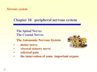
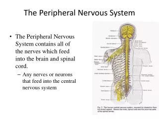
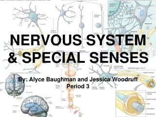
Chapter 6B The Peripheral Nervous System: Special Senses
Outline • Pathways, perceptions, sensations • Receptor Physiology • Receptors have differential sensitivities to various stimuli. • A stimulus alters the receptor’s permeability, leading to a graded receptor potential. • Receptor potentials may initiate action potentials in the afferent neuron. • Receptors may adapt slowly or rapidly to sustained stimulation. • Each somatosensory pathway is “labeled” according to modality and location. • Acuity is influenced by receptive field size and lateral inhibition. • PAIN • Stimulation of nociceptors elicits the perception of pain plus motivational and emotional responses. • The brain has a built-in analgesic system.
Cortex • Higher processing • Basal nuclei • Control of movement, inhibitory, negative • Thalamus • Relay and processing of sensory information • Awareness, a positive screening center for information • Hypothalamus • Hormone secretion, regulation of the internal environment • Cerebellum • Important in balance and in planning and executing voluntary movement • Brain Stem • Relay station (posture and equilibrium), cranial nerves, control centers, reticular integration, sleep control
What did you learn from the vision lab? • Color blindness • Rod and cone function • What an astigmatism is • After imaging? positive and negative after images
Vision outline • Anatomy • Muscles and light control • Refraction and refractive structures • Refractive problems • Retina, photoreceptors, transduction • Visual fields • Visual cortical processing
Countercurrent exchange • Found in many animal systems • thermoregulation, and in the kidney • The transfer of a substance flowing in one direction to another moving in the opposite direction • Efficient - gill can remove 80 % of O2
Eye • Sensory organ for vision • Mechanisms that help protect eyes from injury • Eyeball is sheltered by bony socket in which it is positioned • Eyelids • Act like shutters to protect eye from environmental hazards • Eyelashes • Trap fine, airborne debris such as dust before it can fall into eye • Tears • Continuously produced by lacrimal glands • Lubricate, cleanse, bactericidal
Eye • Spherical, fluid-filled structure enclosed by three tissue layers • Sclera/cornea • Sclera – tough outer layer of connective tissue; forms visible white part of the eye • Cornea – anterior, transparent outer layer through which light rays pass into interior of eye • Choroid/ciliary body/iris • Choroid - middle layer underneath sclera which contains blood vessels that nourish retina • Choroid layer is specialized anteriorly to form ciliary body and iris • Retina • Innermost coat under choroid • Consists of outer pigmented layer and inner nervous-tissue layer • Rods and cones
Eye • Interior consists of two fluid-filled cavities separated by the lens • Posterior cavity • Larger cavity between lens and retina • Contains vitreous humor • Important in maintaining the spherical shape of eyeball • Anterior cavity • Anterior cavity between cornea and lens • Contains aqueous humor • Carries nutrients for cornea and lens • Produced by capillary network within ciliary body
Eye • Iris • Controls amount of light entering eye • Contains two sets of smooth muscle networks • Circular (or constrictor) muscle • Radial (or dilator) muscle • Pigment in iris is responsible for eye color • Unique for each individual • Basis for latest identification technology • Pupil • Round opening through which light enters the eye
Eye • Fovea • Pinhead-sized depression in exact center of retina • Point of most distinct vision • Has only cones • Macula lutea • Area immediately surrounding fovea • Fairly high acuity • Macular degeneration • Leading cause of blindness in western hemisphere • “doughnut” vision
Formation and Drainage of Aqueous Humor Aqueous humor is formed by capillary network in ciliary body, then drains into the canal of Schlemm, and eventually enters the blood.
Vision outline • Anatomy • Light and muscle control • Refraction and refractive structures • Refractive problems • Retina, photoreceptors, transduction • Visual fields • Visual cortical processing
Eye • Convex structures of eye produce convergence of diverging light rays that reach eye
Eye • Two structures most important in eye’s refractive ability are • Cornea • Contributes most extensively to eye’s total refractive ability • Refractive ability remains constant because curvature never changes • Lens • Refractive ability can be adjusted by changing curvature as needed for near or far vision
Eye Focusing on Distant and Near Light Sources What happens to light rays when they leave the light source?
Eye • Accommodation • Change in strength and shape of lens • Accomplished by action of ciliary muscle and suspensory ligaments • Age-related reduction in accommodation ability - presbyopia
Eye • Macula lutea • Area immediately surrounding fovea • Fairly high acuity • Macular degeneration • Leading cause of blindness in western hemisphere • “doughnut” vision
Lasik • Eye Health: LASIK Laser Eye Surgery • Laser in-situ keratomileusis, or LASIK, is a popular surgical approach used to correct vision in people who are nearsighted, farsighted, or have astigmatism. • All laser vision correction surgeries work by reshaping the cornea, or clear front part of the eye, so that light traveling through it is properly focused onto the retina located in the back of the eye. LASIK laser eye surgery (laser in-situ keratomileusis) is one of a number of different surgical techniques used to reshape the cornea. • What Are the Advantages of LASIK Laser Eye Surgery? • LASIK laser eye surgery has many benefits, including: • LASIK laser eye surgery is associated with very little pain. • Vision is corrected nearly immediately or by the next day after LASIK laser eye surgery. • Recovery is quick and usually no bandages or stitches are required after LASIK laser eye surgery. • Adjustments can be made years after LASIK laser eye surgery to further correct vision. • After having LASIK laser eye surgery, most patients no longer need corrective eyewear.
Mechanics of Accommodation Near vision Far vision * Light moves towards thick part of lens
Vision outline • Anatomy • Muscles and light control • Refraction and refractive structures • Refractive problems • Retina, photoreceptors, transduction • Visual fields • Visual cortical processing
Vision outline • Anatomy • Muscles and light control • Refraction and refractive structures • Refractive problems • Retina, photoreceptors, transduction • Visual fields • Visual cortical processing
Eye • Retina – receptor containing portion is actually an extension of the CNS • Neural portion of retina consists of three layers of excitable cells • Outermost layer containing rods and cones • Middle layer of bipolar cells • Inner layer of ganglion cells • Axons of ganglion cells join to form optic nerve • Point on retina at which optic nerve leaves is the optic disc • Region often called the blind spot because no image can be detected here because of lack of rods and cones
Photoreceptors • Rod and cone cells • Consist of three parts • Outer segment • Detects light stimulus • Inner segment • Contains metabolic machinery of cell • Synaptic terminal • Transmits signal generated in photoreceptor on light stimulation to next cells in visual pathway
Photopigments • Undergo chemical alterations when activated by light • Consists of two components • Opsin • Protein that is integral part of disc membrane • Retinene • Derivative of vitamin A • Light-absorbing part of photopigment
Photopigments • Four different photopigments • Rod pigment • Provide vision only in shades of gray • Rhodopsin • Absorbs all visible wavelengths • Cone pigments • Respond selectively to various wavelengths of light • Make color vision possible • Red cones • Green cones • Blue cones
Parasympathetic stimulation Sympathetic stimulation + + Circular (constrictor) muscle runs circularly Circular muscle of iris Radial muscle of iris Pupil Iris Radial (dilator) muscle runs radially Pupillary constriction Pupillary dilation Fig. 6-11, p. 193
The sensitivity of the eyes varies through dark and light adaptation. • Dark adaptation • Can gradually distinguish objects as you enter a dark area. • Due to the regeneration of rod photopigments that had been broken down by previous light exposure. • Light adaptation • Can gradually distinguish objects as you enter an area with more light. • Due to the rapid breakdown of cone photopigments.
Vision outline • Anatomy • Muscles and light control • Refraction and refractive structures • Refractive problems • Retina, photoreceptors, transduction • Visual fields • Visual cortical processing
Hearing outline • Anatomy • Outer, middle, inner • hearing • Transmission of sound waves • Hair cells and transduction • Cochlea and canals/ducts • Pitch and loudness • auditory cortical processing
Ear • Consists of three parts • External ear • Consists of pinna, external auditory meatus, and tympanum • Transmits airborne sound waves to fluid-filled inner ear • Amplifies sound energy • Middle ear • Transmits airborne sound waves to fluid-filled inner ear • Amplifies sound energy • Inner ear • Houses two different sensory systems • Cochlea • Contains receptors for conversion of sound waves into nerve impulses which makes hearing possible • Vestibular apparatus • Necessary for sense of equilibrium
Hearing outline • Anatomy • Outer, middle, inner • hearing • Transmission of sound waves • Hair cells and transduction • Cochlea and canals/ducts • Pitch and loudness • auditory cortical processing
Hearing • Neural perception of sound energy • Involves two aspects • Identification of the sounds (“what”) • Localization of the sounds (“where”) • Sound waves • Traveling vibrations of air • Consist of alternate regions of compression and rarefaction of air molecules
Hearing • Pitch (tone) of sound • Depends on frequency of air waves • Intensity (loudness) • Depends on amplitude of air waves • Timbre (quality) • Determined by overtones