Download
1 / 33
370 likes | 1.04k Vues
Chapter 2 Dental Plaque. Introduction. Dental caries and periodontal diseases are two of the most widespread of all human maladies, resulting from the accumulation of many different species of bacteria Dental plaque is a naturally acquired, multi-species biofilm. FIGURE 2–1

E N D
Chapter 2 Dental Plaque
Introduction • Dental caries and periodontal diseases are two of the most widespread of all human maladies, resulting from the accumulation of many different species of bacteria • Dental plaque is a naturally acquired, multi-species biofilm
FIGURE 2–1 A 13-year-old female with dental caries on the facial surface of the incisors in the maxilla and swollen, discolored gingival tissues around the mandibular incisors, which is characteristic of chronic gingivitis. (Courtesy of Dr. W. K. Grigsby, University of Iowa College of Dentistry, Iowa City.)
FIGURE 2–2 The dental plaque on these teeth has been stained with a discoloring solution and rinsed. Note the presence of plaque interproximally and adjacent to the gingiva, but relatively absent closer to the incis. (Courtesy of Dr. W. K. Grigsby, University of Iowa College of Dentistry, Iowa City.)
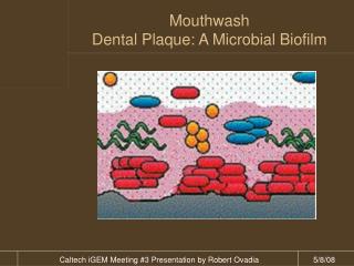
Dental Plaque: Microbial Biofilm Dental plaque is a multi-species biofilm Some bacterial species may be of greater relevance in the development of caries and periodontal diseases Historical aspect: Most natural surfaces have their own coating of microoganisms or biofilm
Historical Aspect One of the first known examples of life is mineralized bacteria or algae attached to rocks from the Precambrian Era (approximately 3800 million years ago) Calculus is a hard calcified deposit of plaque that has become mineralized
Dental Plaque: Biofilm Not all microorganisms within the biofilm population react uniformly to antimicrobial treatment at a given time It is important to include mechanical oral hygiene practices in addition to using antimicrobial therapy to disturb the attached biofilm
Bacterial Colonization of the Mouth Bacteria are acquired from: Atmosphere Food Human contact Contact from animals, such as pets Bacteria subsequently form colonies between saliva and oral soft and hard tissues
Tooth Eruption Prior to eruption the external surface of tooth enamel is lined by remnants of the enamel-forming organ These tissue remnants are the reduced enamel epithelium and the basal lamina This subsurface organic material is called subsurface pellicle
FIGURE 2–3 This transmission electron micrograph demonstrates remnants of the subsurface pellicle (SSP) and the acquired pellicle (AP) between the enamel (ES) surface and the bacterial cells (B) of the dental plaque. (Courtesy of Dr. M. A. Listgarten, University of Pennsylvania School of Dental Medicine, Philadelphia.)
FIGURE 2–4 Junction of reduced enamel epithelium and enamel. The reduced ameloblasts (RA) are attached to the enamel by hemidesmosomes (HD) and a basal lamina (BL). EM, enamel matrix remnants form a subsurface pellicle; ES, enamel space. Original magnification × 45,000. (Courtesy of Dr. M. A Listgarten, University of Pennsylvania School of Dental Medicine, Philadelphia.)
Tooth Eruption (Continued) An erupted tooth immediately becomes covered by a thin, microscopic coating of saliva materials It is acquired after tooth eruption and is exogenous in nature Referred to as acquired pellicle
The Acquired Pellicle Acellular Consists primarily of glycoproteins derived from saliva Occupies million of microscopic voids in the erupted tooth caused by chemical and mechanical interactions of the tooth surface with the oral environment If the pellicle is displaced by a prophylaxis it begins to reform immediately
The Acquired Pellicle (Continued) Also forms on dental restorations Interestingly, there is competition for binding sites on the pellicles, not only by receptors on bacteria but also from host proteins, including immunoglobulins
Dental Plaque Formation Mechanical displacement Stagnation Availability of nutrients
FIGURE 2–5 Scanning electron micrograph of dome formation in the plaque. (From Brady, J. M. (1973). J Periodontol, 44:416–28.)
Molecular Mechanisms of Bacterial Adhesion • The initial bacterial attachment to the acquired pellicile is thought to involve physicochemical interactions between molecules or portions of molecules • Bacteria have external cell-surface proteins termed adhesions
FIGURE 2–6 This diagram illustrates some of the possible molecular mechanisms that mediate attachment of bacteria to teeth during dental plaque formation. A. A side chain of a phenylalanine component of a bacterial protein interacts via hydrophobic bonding with a side chain of a leucine component of a salivary glycoprotein in the acquired pellicle. B. The negatively charged carboxyl group of a bacterial protein is attracted to a positively charged calcium ion (i.e., electrostatic attraction), which in turn is attracted to a negatively charged phosphate group of a salivary phosphoprotein in the acquired pellicle. C. The host’s dietary sucrose is converted by the bacterial enzyme, glucosyltransferase, to the extracellular polysaccharide, glucan, which has many hydrophobic groups and can interact with amino acid side-chain groups, such as serine, tyrosine, and threonine. D. The fimbrial surface appendage extends from the bacterial cell to permit the terminal adhesin portion to bind to a sugar component of a salivary glycoprotein in the acquired pellicle.
Bacteria in the Dental Plaque Supragingival Subgingival Primary colonizers Secondary colonizers
Dental Plaque Matrix • Dental plaque consists of different species of bacteria that are not uniformly distributed • The bacterial cells in the biofilm are surrounded by an intercellular plaque matrix which is composed of both organic and inorganic components
FIGURE 2–7 An electron micrograph showing palisades (P) of bacteria perpendicular to the enamel surface (ES), bacterial cells that are probably secondary colonizers (SC), the intercellular plaque matrix (IPM), and the acquired pellicle (AP). (Courtesy of Dr. M. A Listgarten, University of Pennsylvania School of Dental Medicine, Philadelphia.)
FIGURE 2–8 A. Cross section of “corn cob” from 2-month-old plaque. A coarse fibrillar material attaches the cocci (C) to the central filament (CF). Original magnification × 22,500. (From Listgarten, M. A., Mayo, H. E., & Tremblay, R. (1975). J Periodontol, 46:10–26.) B. Coarse “test-tube brush” formations consisting of central filament (CF) surrounded by large, filamentous bacteria with flagella uniformly distributed over its body (LF). Background consists of a spirochete-rich microbiota (S). Original magnification × 4,300. (From Listgarten, M. A. (1976). J Periodontol, 47: 1–18.)
Dental Plaque Metabolism • A source of energy is required for metabolism to occur • Once exposed to sucrose, microorganisms produce: • Acid • Intracellular polysaccharides • Extracellular polysaccharides
Dental Calculus • The term calculus is derived from the Latin word meaning pebble or stone • Calculus itself is not harmful and cannot be removed by brushing or flossing • It needs to be removed because its presence makes routine oral hygiene difficult • May be affected by behavioral factors and systemic conditions
FIGURE 2–9 Radiograph demonstrating a “spur”- shaped deposit of calculus (C) on the distal side of the left first molar in the maxilla. The calculus is apical to the overhanging metallic restoration (R). The arrow (G) marks the coronal level of the gingival tissues, which indicates that this is a subgingival deposit of calculus. (Courtesy of Dr. W. K. Grigsby, University of Iowa College of Dentistry, Iowa City.)
Dental Calculus (Continued) • Crystals in calculus include: • Hydroxyapatite • Brushits • Whitlockite
FIGURE 2–10 Deposits of supragingival calculus on the lingual surface of incisors and canines that could not be removed by brushing. (Courtesy of Dr. W. K. Grigsby, University of Iowa College of Dentistry, Iowa City.)
Dental Calculus (Continued) • The two types of calculus may differ in color: • Supragingival calculus usually appears as a yellow to white mass with a chalky consistency • Subgingical calculus appears gray to black in color and has a flint-like consistency
FIGURE 2–11 Typical pattern of dental plaque mineralization in which the initial mineralization occurs in the inter-bacterial plaque matrix (M), with bacterial cells (B) becoming mineralized secondarily. Original magnification × 40,000. (Courtesy of Dr. M. A. Listgarten, University of Pennsylvania School of Dental Medicine, Philadelphia.)
FIGURE 2–12 Atypical pattern of dental plaque mineralization in which bacterial cells (B) act as foci of initial mineralization, with the matrix (NM), becoming mineralized secondarily. Original magnification × 25,000. (Courtesy of Dr. M. A. Listgarten, University of Pennsylvania School of Dental Medicine, Philadelphia.)
Dental Calculus (Continued) Attachment of calculus to the teeth Inhibition of calculus formation
Summary Microbial film Bacterial colonization Acquired pellicle Dental plaque formation Molecular mechanisms
Summary (Continued) Bacteria in the dental plaque Dental plaque matrix Dental plaque metabolism Dental calculus