MODULE V
470 likes | 484 Vues
This module discusses the main causes of death in children following a disaster and the IMCI strategy for managing prevalent infections. It includes guidelines for classification and management based on the child's age, as well as information on danger signs and antibiotic arsenal. The module also provides insights into influenza and measles infections in children.

MODULE V
E N D
Presentation Transcript
MODULE V Management of Prevalent Infections in Children Following a Disaster
MAIN CAUSES OF DEATH • Acute respiratory infections • Diarrhea and dehydration • Measles • Malaria • Malnutrition
The IMCI strategy 2 components based on the child’s age: • sick young infant aged up to 2 months • sick child aged 2 months up to 5 years
The IMCI strategy • The clinical decision making approach involves using a limited number of symptoms and signs to classify the severity of illness, which determines the management with guidelines for follow-up, counseling for the parents, and instructions regarding when to return additional care is needed.
Management • Pink: needs to be urgently referred to a higher level of care • Yellow: requires specific treatments • Green: can be safely managed at home with supportive care
Sick young infant aged up to 2 months • Classification and management of severe disease (pneumonia, meningitis, and sepsis), local bacterial infection, jaundice, diarrhea, HIV infection, poor weight gain, breast feeding and other feeding problems, immunization status, and mother’s health.
Severe disease (PINK) • Not feeding, convulsions, fast breathing (more than 60 breaths per minute) severe chest indrawing, fever or low temperature, and lack of movement. • Refer urgently to the hospital with a first antibiotic dose and treatment to prevent low blood sugar
Local bacterial infection (YELLOW) • Signs of umbilical infection (redness and or purulent discharge) or skin pustules • Treat with an appropriate antibiotic.
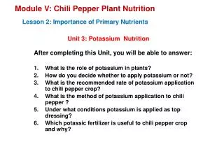
Sick child aged 2 months up to 5 years • Classification and management of respiratory disease, diarrhea, febrile illness (malaria), measles, ear infections, malnutrition, anemia, HIV, and immunization status.
IMCI STRATEGY DANGER SIGNS Unable to drink or breast feed (too weak) Vomits everything Had convulsions Lethargic or unconscious Convulsing now
IMCI: COUGH OR DIFFICULT BREATHING Very severe respiratory disease Any general danger sign Stridor in a calm child Pneumonia Fast breathing Chest indrawing Cough without pneumonia No signs of pneumonia or severe disease
ANTIBIOTIC ARSENAL Oral antibiotics Amoxicillin Cotrimoxazole (TMPSMX) Intramuscular (IM) antibiotics Benzylpenicillin Cefuroxime or Ceftriaxone
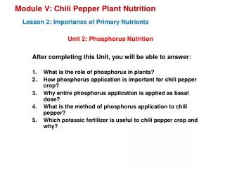
INFLUENZA VIRUS • Family Orthomyxoviridae • “myxo” mucus • segmented, single-stranded RNA • Influenza A first isolated 1933; Influenza B 1940 • 15 hemagglutinin (HA) and 9 neuraminidase (NA) subtypes • Only H1N1, H2N2, H3N2 subtypes associated with widespread epidemics in humans
Type A Potentially severe illness Epidemics and pandemics Rapidly changing Birds, swine, horses, seals, humans Type B Usually less severe illness Epidemics More uniform Humans Type C Usually mild or asymptomatic illness Minimal public health impact Humans, swine CLINICALLY RELEVANT INFLUENZA VIRUSES
INFLUENZA: A CONTINUOUSLY CHANGING VIRUS Hemagglutinin (HA) *cell entry Neuraminidase (NA) *cell escape M1, M2 Nucleoprotein (NP) Polymerase Proteins (PP) Adapted from: Hayden FG et al. Clin Virol. 1997:911-942.
RNA Hemagglutinin Neuraminidase Antibodies Sialic acid ANTIGENIC DRIFT (A & B)
TRANSMISSION OF INFLUENZA • Person to person • Droplet spread • small particle aerosols • Fomite contamination • Steel and plastic 24-48 hrs • Cloth, paper, tissues 8-12 hrs • Hands 5 min (high viral titer) • Principal site of replication- columnar epithelium • Incubation period- 18 hrs to 5 or more days (average 2-3 days) • Virus shedding 3-7 days • Viral titers are generally higher in young children with shedding lasting 10 days or longer
RECOGNIZING PEDIATRIC INFLUENZA Neonates Infants/Toddlers Children/Teens High fever GI symptoms Rapid onset Lethargy Fever >103°F (>39.5°C) High fever Decreased eating Anorexia Cough Mottling Respiratory syndromes Chills Apnea Malaise Headache Sore throat
Acute otitis media (children) Sinusitis Pneumonia Exacerbation of underlying illness Dehydration (infants) Encephalopathy Reye syndrome (children) Myositis Myocarditis Febrile seizures INFLUENZA VIRUS INFECTION COMPLICATIONS Common Complications Uncommon Complications
MEASLES Highly contagious infection (98-100% in susceptible contacts) Transmission through respiratory secretions (contact and aerosolized particles) Incubation period: 10-14 days Mortality rate Nutrition / crowding / inoculum Overcrowded living conditions are an important triggering factor for epidemics
NATURAL HISTORY OF MEASLES Incubation 10-14 days Day 1 Day 2 Day 3 Day 4 Day 5 Day 6 Day 7 Fever ------------- FEVER--------------------] Exposure Cough ---------------------------------------------------- - - - Conjunctivitis ----------------------------------------- - - - Coryza ------------------------------------------------- - - - Köplik spots---] Rash ------------------- Identification of one case in a camp should speed up immunization process
MEASLES AND VITAMIN A DEFICIENCY MEASLES unmasks an underlying Vitamin A deficiency SYNERGIC EFFECT VITAMIN A DEFICIENCY (even subclinical) increases measles-associated morbidity and mortality Measles-associated morbidity and mortality may be reduced by administering Vitamin A to high risk populations
Measles Managment • Evaluate for associated infections • Classify any child having a general danger sign, clouding of the cornea, or deep or extensive mouth ulcers as severe complicated measles and refer urgently to the hospital with vitamin A, the first dose of an appropriate antibiotic, and if there is eye discharge or corneal clouding an dose of tetracycline eye ointment.
Measles Managment • The presence of eye drainage and or mouth ulcers without other signs is classified as yellow. Treatment includes Vitamin A, tetracycline eye ointment for eye discharge, and gentian violet for mouth ulcers. These children need a follow up visit in 3 days. • A child without complications is green and needs only vitamin A.
ALGORITHM FOR A SUSPECTED CASE OF MEASLES Child with fever and rash consistent with measles Report case to Alert System Case Confirmation • Laboratory tests Local response • Guarantee vaccines • Vitamin A • National Response Team Search for other cases and Quarantine Start response and prevention Measles vaccine Priority groups Resources and logistics
DENGUE Incubation Abdominal Pain Cyanosis Shock Hemorrhages Hepatitis Plasma leakage Headache Myalgia Rash Bone pain Vomiting
Grade Hemorrhage Platelets Capillary Permeability I Positive <100,000 Plasma leakage* tourniquet test II Spontaneous <100,000 Plasma leakage* bleeding III (DSS) Spontaneous <100,000 Plasma leakage+ bleeding PP <20 mmHg Hypotension IV (DSS) Spontaneous <100,000 Profound shock bleeding Absent pulse or BP WHO GUIDELINES FOR THE DIAGNOSIS OF DENGUE HEMORRHAGIC FEVER (DHF) *Hct admission >20%/age or reduction Hct >20% post-resuscitation fluids PP: pulse pressure
80% asymptomaticinfections Unusual manifestations Hepatitis Encephalopathy Pancreatitis Pleural effusion DENGUE MANIFESTATIONS IN CHILDREN
Rest Acetaminophen/Paracetamol No aspirin or NSAIDs No antibiotics Oral rehydration (WHO solution) 50 mL/kg over 4-6 hours Maintenance 80-100 mL/kg/day Monitor CNS signs MANAGEMENT OF THE CHILD WITH DENGUE
Hospitalization in case of grade II HDF Platelets <100,000 Hematocrit > 20% over normal Colloid solutions at 6 mL/kg/hr MANAGEMENT OF THE CHILD WITH HEMORRHAGIC DENGUE Improvement Worsening 3 mL/kg/hr 10 mL/kg/hr
MALARIA Caused by a protozoal blood parasite capable of causing a wide spectrum of diseases Plasmodium vivax Plasmodium ovale Plasmodium malariae Geographical distribution:Tropic / Subtropics Transmission: Anopheles mosquito Plasmodium falciparum
MALARIA SUSCEPTIBILITY In endemic areas, there is partial immunity in older children and adults due to previous infection Most susceptible individuals to severe and fatal malaria: Non-immune and immunocompromised people Infants and young children, pregnant women and malnourished Plasmodium falciparum-infected people Infection Identification of parasitemia Asymptomatic Disease Presence of signs and symptoms Acute, subacute, chronic
FEVER 39 39 38 38 37 37 Non-specific Pattern Classical Pattern MALARIA CLINICAL MANIFESTATIONS Partially immune patients may develop moderate fever with a non-specific pattern Patients will feel and look sick due to fever, but they will feel relatively well between paroxysms of fever Associated chills, headache, myalgia
Severe Malaria • Parasitemiais >5% • Any of the following complications: -prostration (patient unable to sit or walk) -multiple convulsions -impaired consciousness not attributable to another cause -abnormal bleeding -meningeal signs -jaundice ( hemolysis)
Malaria Diagnosis • Rapid diagnostic tests • Bedside testing • Thick and thin blood smears • Difficult in a disaster situation
Malaria Management • The clinical diagnosis of malaria based on non specific signs and symptoms tends to be highly inaccurate. • When a patient presents with febrile illness who lives in an area with malaria, in the absence of available diagnostic testing begin treatment when the clinical history and presentation are consistent with malaria.
Types of Malaria P. falciparum– Most severe type of MALARIA (MALIGNANT) High lethality rate in infected individuals Highly drug-resistant Plasmodium vivax “BENIGN” MALARIA Plasmodium ovale Most are sensitive Plasmodium malariae to chloroquine These infections cause morbidity and contribute to multifactorial mortality
Treatment of Uncomplicated Malaria: P. Falciparum or Unknown Species Preferred Therapies (check your country policy): Atovaquone-Proguanil (Malarone) 4 adult tabs (1000mg Atovaquone) po qd x 3 days Artemether-lumefantrine (Coartem) 4 tablets immediately, 4 tablets 8 hours later, then 4 tablets BID for 4 more doses Second-Line Therapies: Quinine sulfate plus: Doxycycline, Tetracycline, or Clindamycin Mefloquine
Uncomplicated Malaria: Chloroquine-Sensitive Species/Areas Children: a total dose of 25 mg/kg of CHLOROQUINE over a 3-day period t = 0 10 mg/kg po t = 6 h 5 mg/kg po or 10mg/kg t = 24 h 5 mg/kg po at t = 24 h t = 48 h 5 mg/kg po Adults: similar schedule. 1 gr followed by 500 mg x 3 Pregnant women: Malaria is SEVERE. Chloroquine treatment is safe
Malaria Supportive Treatment Fever control Antipyretics, no more than a few doses Cool compresses Dehydration Oral rehydration solution, increased need for fluids Malnutrition Assess and treat Anticipate symptom resolution at 48-72 hours
Severe complicated malaria treatment • First line (preferred treatment) is Artesunateparentral (IV/IM). • In the absence of parenteral form of Artesunate, Artemether IM is acceptable. • Quinine is acceptable option but requires attention to the proper dosage and administration with IV fluids. There is a loading dose and maintenance dose and care needs to be taken to prevent hypoglycemia