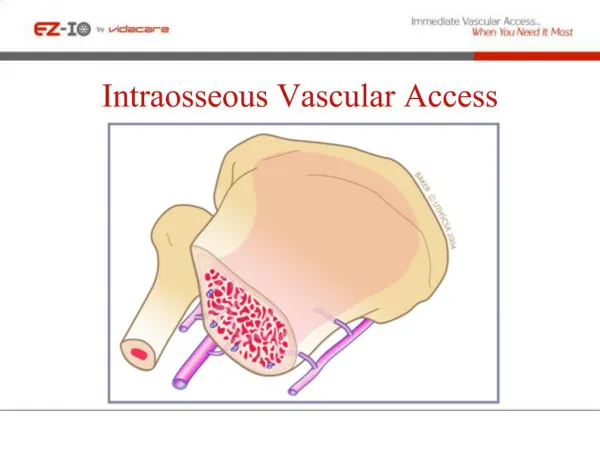
Vascular Access How I Do It
Vascular Access How I Do It. Gareth Griffiths Department of Vascular Surgery, Ninewells Hospital, Dundee, UK Chairman of the Specialty Advisory Committee in General Surgery. Team Working. Nephrologist Vascular technician Dialysis nurses Radiologist Anaesthetist Surgeon.

Vascular Access How I Do It
E N D
Presentation Transcript
Vascular Access How I Do It Gareth Griffiths Department of Vascular Surgery, Ninewells Hospital, Dundee, UK Chairman of the Specialty Advisory Committee in General Surgery
Team Working • Nephrologist • Vascular technician • Dialysis nurses • Radiologist • Anaesthetist • Surgeon
Pre Operative Assessment • Clinical • Ultrasound • Vein • Size • Intravenous webs / thrombus • Beware of spasm • Examine full length • Central deep veins • Artery • Wall calcification • Waveform pattern • Examine full length
Pre Operative Assessment • Suspicion of central vein stenosis • Venography • MR • CT • Catheter
Pre Operative Assessment • Upper limb before lower limb • Non-dominant before dominant • Distal before proximal • Autogenous before prosthetic • Priority depends on the patient • Already on haemodialysis • Age
Sequence of Operations • Radio-cephalic • Brachio-cephalic • Brachio-basilic • Brachio-axillary PTFE • Long saphenous thigh straight / loop • PTFE thigh straight / loop • Superficial femoral vein straight / loop
Rarely Needed Options • Iliac artery – vein PTFE loop • Necklace axillary artery – vein PTFE • Arterio-arterial PTFE
Operative Technique • Local or regional anaesthesia • Gentle handling of tissues • Meticulous technique • Microvascular instruments • Magnification and light
Operative Technique • Radiocephalic / brachiocephalic • Single incision when possible • Mobilise vein • Avoid twisting • Isolate artery • Microvascular clamps • Careful siting of arteriotomy • Relieve spasm – hydrostatic pressure, balloon • Check, check, check
Operative Technique • Brachiobasilic • One stage procedure • Mobilise maximum length of vein • 3-4 short incisions ?endoscopic • Fashion tunnel to match vein length • Straight tunnel preferable • Arched tunnel if vein short
Operative Technique • Prosthetic fistula • 6mm PTFE • Miller cuff at each end • Protects native vessels from PTFE thrombosis • Facilitates removal of infected PTFE
Follow Up • Life long surveillance • Clinical • Bleeding • Cannulation difficulties • Ultrasound • Duplex identified stenosis • Dialysis parameters • Venous pressure < 180mm Hg • Arterial pressure > -180 mmHg • Urea reduction ratio >70% • Access flow >600ml/min <25% fall
Multidisciplinary Meeting • Surgeon • Vascular technologist • Nephrologist • Radiologist • Dialysis specialist nurse • Discuss all patients with duplex or dialysis identified issues • Review all post intervention outcomes
Multidisciplinary Meeting • Selective intervention • Dialysis parameter abnormality + identified stenosis • Angioplasty first • Cutting balloon if necessary • Stenting if necessary • Surgical re-fashioning • Failed endovascular intervention
Multidisciplinary Meeting • Surveillance and repeated intervention • Longest assisted primary patency possible • Pre-emptive new fistula creation • When fistula failure is predicted • Before loosing fistula access
Fistula Thrombosis • Attempt salvage unless • Fistula had been identified as failing • Active infection • Aneurysmal with organised thrombus • Radiological salvage • Combined mechanical and lytic • Concomitant angioplasty / stenting when needed • Surgical thrombectomy • Early post op thrombosis
Aneurysmal Fistulae • No issue if uncomplicated • Cosmetic • Ask patient to accept • Thin skin • Lateral cannulation • Bleeding • Repair with vein buttress occasionally possible • Ligation often needed
Steal • Exclude central arterial stenosis • Often mild • Conservative management • Significant • Pain, tissue loss • High flow • Good distal vessels • High fistula flow • Low flow • Diseased distal vessels • Critical flow distally
Steal • High flow • Assess direction of flow in distal artery • If retrograde • Radial fistula – ligate distal radial artery • Maximises ulnar flow into hand • Brachial fistula - Distal Revascularisation and Interval Ligation (DRIL) • Restores antegrade flow towards hand • If antegrade • Band fistula • Reduces fistula flow, improves distal perfusion
Steal • Low flow • Diseased distal vessels • Poor outlook • Increasingly common • Banding • Rarely possible – fistula flow already low • Ligate fistula
Swollen Arm • Assess promptly • Generally indicates central vein stenosis • Urgent catheter venography • Angioplasty or stenting when possible • May need fistula ligation • Try to avoid with careful pre-op assessment
Vascular AccessHow I Do It • Multi-disciplinary team • Attention to detail • Logical sequence for fistula creation • Perseverance • Fistula surveillance and repeated interventions