Download
1 / 85
860 likes | 1.2k Vues
Treatment of Lymphedema. Lymph Anatomy. Lymph nodes Lymph vessels Thymus gland Spleen Tonsils Peyer’s patches. Lymph Vessels. Capillaries Pre-collectors Collectors Trunks. Lymph Capillaries. Larger diameter than blood capillaries No valves Lymph can flow in any direction

E N D
Lymph Anatomy • Lymph nodes • Lymph vessels • Thymus gland • Spleen • Tonsils • Peyer’s patches
Lymph Vessels • Capillaries • Pre-collectors • Collectors • Trunks
Lymph Capillaries • Larger diameter than blood capillaries • No valves • Lymph can flow in any direction • Can absorb interstitial fluid
Pre-Collectors & Collectors • Pre-Collectors • Channel lymph fluid into transporting vessels • Can absorb fluid • Collectors • Transporters • Resemble veins in structure • Passive valves: ever .6-2cm along vessel • Lymphangioactivity • Contractions caused by Sympathetic Nervous System and lymph volume • Superficial and deep
Trunks & Ducts • Largest lymph vessels • Thoracic duct-largest, pumping by the diaphram. • From in cisterna chyle • Ducts empty into venous system
Lymph Fluid/Lymphatic Load • Consists of: • Proteins (1/2 of bodies protien) • Water • Cells (RBC, WBC, Lymphocytes) • Waste Products • Fat (intestinal lymph, chyle)
Lymph Nodes • Filtering station for bacteria, toxins, & dead cells • Produces lymphocytes • Regulates the concentration of protein in the lymph • Typically thickens the fluid • 600-700 in body
Lymphatic Watersheds • Median-Sagittal • Tranverse • Clavical • Spine of Scapula • Chaps or Gluteal
Lymph Time Volume & Transport Capacity • LTV= amount of lymph which is transported by the lymphatic system in a unit of time • TC=maximum lymph time volume • Functional Reserve=the difference between the LTV and the TC
Defining Types of Lymphatic Insufficiencies High Volume or Dynamic Insufficiency Low Volume or Mechanical Insufficiency • High Output Failure • Leads to Edema • Low Output Failure= Lymphedema TC TC LL=LTV LL LTV
Lymph Propulsion • Arterial pulsation • Muscle pump • Respiration • Contraction of the lymphangion
Definition of Lymphedema • Lymphedema is the result of the abnormal accumulation of protein rich edema fluid • Primary or secondary • Afflicts approximately 1% of the US population (2.5 million people) * A SUDDEN ONSET OF EDEMA MUST BE THOROUGHLY EVALUATED BY A PHYSICIAN
Physical Exam • History • Inspection • Measurements: weight, circumference • Skin assessment: nodules, bumps, discoleration • Palpation • Temperature: usually a bit warmer • Stemmer’s sign: rolls on finger, square and thick skin • Skin fold(s) • Pitting • Fibrosis
Other Diagnostic Tests • Lymphography • Venous Doppler or Venous Sonography • Indirect Lymphography • Fluorescence Microlymphography • Lymphoscintigraphy • CT Scan • MRI
Types of Lymphedema Primary Secondary • Hypoplasia (not as many lymph nodes) • Hyperplasia • Aplasia • Inguinal Node Fibrosis • (Kineley Syndrome • Milroy’s Disease-congentital, males, unilateral typically • Meige’s Syndrome: most females around puberty, Bilateral, webbing of fingers and toes, two rows of lashes • Surgery • Radiation Therapy • Trauma: blunt trauma • Filariasis: parasite, blocks lymph nodes • Cancer (Malignant) • Infection • Obesity • Self Induced
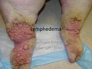
Stages of Lymphedema • Latency Stage • Reduced transport capacity • No noticeable edema • Stage I • Pitting edema • Edema reduces with elevation (no fibrosis) • Tight sleeve during the day • Stage II • Pitting becomes progressively more difficult • Connective tissue proliferation (fibrosis) • Stage III • Non pitting • Fibrosis and Sclerosis • Skin changes (papillomas, hyperkeratosis, etc)
Differential Diagnosis • Lipidema: females, symmetrical (no feet), no pitting, very painful to palpations, bruise easily, tissue is softer. • Chronic Venous Insufficiency: gaiter distribution, non-pitting, hemosiderin staining, fibrotic. • Acute Deep Venous Thrombophlebitis: swelling, redness, painful, sudden onset • Cardiac Edema: bilateral, pitting, complete resolution when legs elevate above heart, no pain. • Congestive Heart Failure: pitting, dyspnea, jugular vein distention. • Malignancy: • Filariasis: • Myxedema: decreased ability to sweat, orange skin • Complex Regional Pain Syndrome (RSD, Sudeck’s)
Lymphedema Interventions • Surgery (Debulking, Liposuction) • Taking out all the lymphatic with these surgeries • Medication (Diuretics, Benzopyrones) • Takes out all the water, but leaves lymphatic's with protein rich lymph fluid. • Pneumatic Compression Pump • May harden the tissue or destroy lymph collectors, and leave person immobile for a couple of hours. • COMPLETE DECONGESTIVE THERAPY • Removes proteins from the system.
Anti-Edema Medications • Not effective because: • Do not allow the proteins to be reabsorbed into the venous system • As long as proteins are stagnate in the interstitial space the onconic pressure remains high and lymphedema persists • Can worsen Lymphedema in the long run as they increase the concentration of proteins in the interstitial space exacerbating fibrosis
Treatment Schools of Thought • Casley-Smith • Foldi • LeDuc • Vodder • Norton • Klose
Complete Decongestive Therapy (CDT) • Skin Care • Manual Lymph Drainage • Compression Therapy • Remedial exercise
Purpose of lymphatic treatment • Applied pressure softens fibrotic tissue • Excess protein is removed • Formation of new tissue channels through anastomoses • Provide support • Enhance oxygenation by decongesting areas where lymph volume is high • Long-term maintenance of improved limb size and shape
Contraindications (precautions) to CDT • Acute bacterial or viral infection • Wait 24 hours of antibiotic treatment before resuming treatment. • Acute CHF • h/o CHF treat conservative, 1 limb at a time • Kidney malfunction • Untreated malignancy • The existence of impaired arterial perfusion for compression • ABI < 0.50
Patient education • Protect the skin • Signs of infection • Gradual return to activity • Self management • Self massage • Compression garments • Exercises • Weight Management • Obesity and body fluid volume fluctuations are beginning to be associated with the development of lymphedema
Protect the skin : Individuals that have had lymph nodes removed are at risk for lymphedema. To minimize this risk the following precautions should be followed: • Keep arm clean and dry. • Apply moisturizer daily to prevent chapping/chaffing of the skin. • Balance lotion • Attention to nail care; do not cut cuticles. • Protected exposed skin with sunscreen and insect repellent. • Use care with razors to avoid nicks and skin irritation. • Avoid punctures such as injections and blood draws.
Wear gloves while doing activities that may cause skin injury If scratches/punctures to skin occur, keep clean and observe for signs of infection. Gradually build up the duration and intensity of any activity or exercise, and monitor arm during and after for any change in size, shape, firmness or heaviness. Avoid arm constriction from blood pressure cuffs, jewelry and clothing Avoid prolonged (>15 minutes) exposure to heat, particularly hot tubs and saunas Airplane flights: due to decrease pressure in cabin, will need a compression sleeve
Signs of infection • Red • Hot • Pain • Swelling • Fever • Generalized Fatigue
Exercises • Effect of movement on lymphatics - lymph flow; abdominal breathing • Development of an effective exercise program 1.) flexibility exercises 2.) strengthening exercises 3.) aerobic exercises 4.) response of limb is important
Lymphatic Drainage Exercises • Move fluids through lymphatic channels • Active repetitive ROM exercises are performed • Follow a specific sequence to move lymph away from a congested area • Proximal to distal • Avoid static dependent postures
Lymphatic Drainage Exercises • 20 – 30 minutes each session • Twice daily • 7 days a week • Wear compression bandages or garment during exercises • Combine with deep breathing • Rest if possible for 30 minutes following exercises • Check for redness or increased swelling
Sequence of exercises • Proximal starting at neck and trunk • Proximal joints moving distally • 5 reps – 20 reps
Manual Lymph Drainage (MLD) a manual technique to mobilize fluid in the lymph system, by movement of proteins and fluid into the initial lymphatic vessels. This manual technique is done lightly and slowly.
Manual Lymph Drainage (MLD) Basic Principles: • 1. Proximal area is treated first, clearing first the adjacent and unaffected lymphotomes, then proximal sections of the affected lymphotomes. • 2. The direction of pressure depends on the areas of edema and the direction should always be towards a cleared lymphotome. • 3. Technique and variations are repeated rhythmically. • 4. Pressure phase lasts longer than relaxation phase. • 5. As a rule there should be no reddening of the skin
Manual Lymph Drainage (MLD) Techniques: • 1. Call-up - proximal to edema • To clear the collectors proximal to the area • Using the Thumb side of hand • 2. Reabsorbtion - edematous region • Using the 5th digit side of hand • Increases protein reabsorption
Manual Lymph Drainage (MLD) • 1. Mobilize the skin • 2. Apply Pressure • 3. Relax • Technique is done lightly and slowly
MLD – Upper extremity • 1: Supraclavicular nodes • 2: Axillary nodes • 3: Inguinal nodes • 4: Thigh • 5: Popliteal fossa • 6: Calf • 7: Malleolli • 8: Dorsum of foot • 9: Toes
MLD – Upper extremity • 1: Supraclavicular nodes • 2: Axillary nodes • 3: Anterior chest • 4: Back • 5: Mascagni Pathway • 6: Upper arm • 7: Cubital nodes medial/lateral elbow • 8: Forearm supination / pronation • 9: Dorsum/palm of hand • 10: Fingers