Shigella
Shigella. Dr.T.V.Rao MD. Shigella a Highly Infectious Bacteria. Shigella is one of the most infectious of bacteria and ingestion of as few as 100-200 organisms will cause disease.

Shigella
E N D
Presentation Transcript
Shigella Dr.T.V.Rao MD Dr.T.V.Rao MD
Shigella a Highly Infectious Bacteria • Shigella is one of the most infectious of bacteria and ingestion of as few as 100-200 organisms will cause disease. • Most individuals are infected with shigellae when they ingest food or water contaminated with human fecal material. • Shigella can survive up to 30 days in milk, eggs, cheese or shrimps. Dr.T.V.Rao MD
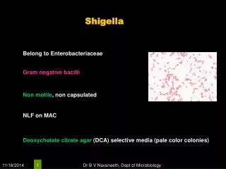
Definition • An enterobacteriaceae • Gram negative bacilli. • Readily growth O2 + An O2. • Metabolically active, fermenting a variety of substrates. • Mostly non-motile, non sporing, non acid fast, 2-4um x0.4 -0.6um rounded ends. Dr.T.V.Rao MD
CLASSIFICATION • 4 SPECIES/SUBGROUPS BASED ON BIOCHEMICAL AND SEROLOGICAL CHARACTERS • SHIGELLA DYSENTERIAE : 12 Serotypes • SHIGELLA FLEXNERI : 6 serotypes • SHIGELLA BOYDII : 18 • SHIGELLA SONNEI : 17 Colicins types Dr.T.V.Rao MD
Taxonomy Family Enterobacteriaceae • Shigella dysenteriae: most serious form of bacillary dysentery • Shigella flexneri: shigellosis in underdeveloped countries • Shigella sonnei: shigellosis in developed countries • Shigella boydii Dr.T.V.Rao MD
Morphology & Physiology • Small Gram-negative, facultatively anaerobic, coliform bacillus • Non-motile (no H antigen) • Possess capsule (K antigen) and O antigen • K antigen not useful in serologic typing, but can interfere with O antigen determination • O antigens: A, B, C, D correspond respectively to the four species • Non-lactose fermenting • Bile salts resistant: trait useful for selective media ferment glucose Reduce nitrates (NO3 to NO2 or N2) are oxidase negative Dr.T.V.Rao MD
CLASSIFICATIONon Basis of Mannitol Fermentation: • 1. Non-mannitol-fermenters • Shigella dysenteria • 2. Mannitol-fermenters • Shigella flexneri • Shigella boydii • Shigella sonnei Dr.T.V.Rao MD
MORPHOLOGY AND STAINING: • Short rods • - Non-encapsulated • - Non-motile • - Non-spore former • - Gram-negative Dr.T.V.Rao MD
CULTURAL CHARACTERISTICS All members of Shigella are aerobic and facultative anaerobes. Grow readily in culture media at pH 6.4 to 7.8 at 10 oC - 40 oC, with optimum of 37 oC. After 24 hours incubation, Shigella colonies reaches a diameter of about 2 mm. The colonies are circular, convex, colorless, but moderately translucent with smooth surface, and entire edges. Dr.T.V.Rao MD
Growth on Selective Medium • In XLD they appear pinkish to reddish colonies while in Heaktoen Enteric Agar (HEA), they give green to blue green colonies. Dr.T.V.Rao MD
HABITAT AND TRANSMISSION Shigella species are found only in the human intestinal tract. Carriers of pathogenic strains can excrete the organism up to two weeks after infection and occasionally for longer periods. Shigella are killed by drying. Shigella are transmitted by the fecal-oral rout. The highest incidence of Shigellosis occur in areas of poor sanitation and where water supplies are polluted. Dr.T.V.Rao MD
Factors Contributing Spread • Spread is always from a human resource and generally involves one of the five f`s: • food, • fingers, • feces, • flies or • fomites. • This is in contrast to salmonellae, which are often spread to humans from infected animals. Dr.T.V.Rao MD
PATHOGENESIS • SOURCE : MAN: CASE OR CARRIER • MODE OF SPREAD: CONTAMINATED FINGERS, FOOD, FLIES, FOMITES • PERSON TO PERSON TRANSMISSION • INFECTIVE DOSE: 10-100 VIABLE BACILLI • HIGHEST CONCENTRATION IN STOOL DURING EARLY/ACUTE INFECTION 103 TO 109 VIABLE BACILLI PER GRAM OF STOOL • POST CONVALESCENT SHEDDING : LOW COUNTS 102 TO 103 Dr.T.V.Rao MD
Transmission • Faecal-oral transmission is the main path of Shigella infection. Other modes of transmission include ingestion of contaminated food or water, contact with infected objects, or sexual contact. Outbreaks of Shigella infection are common in places where sanitation is poor. Dr.T.V.Rao MD
Pathogenesis and Virulence Factors (cont.) Invasiveness in Shigella-Associated Dysentery • Penetrate through mucosal surface of colon (colonic mucosa) and invade and multiply in the colonic epithelium but do not typically invade beyond the epithelium into the lamina propria (thin layer of fibrous connective tissue immediately beneath the surface epithelium of mucous membranes) • Preferentially attach to and invade into M cells in Peyer’s patches (lymphoid tissue, i.e., lymphatic system) of small intestine Dr.T.V.Rao MD
PATHOGENIC DETERMINANTS O antigen: The ability to survive the passage through the host defenses may be due to O antigen. Invasiveness:Virulent Shigella penetrate the mucosa and epithelial cells of the colon in an uneven manner. Intracellular multiplication leads to invasion of adjacent cells, inflammation and cell death. Cell death is probably due to cytotoxic properties of shiga toxin that interfere with protein synthesis. The cellular death and resulting phagocytosis response by the host accounts for the bloody discharge of mucus and pus and shallow ulcers characteristic of the disease. Other toxins: It has a protein toxin which may be neurotoxic, cytotoxic, and enterotoxic. The enterotoxic property is responsible for watery diarrhea. Dr.T.V.Rao MD
PATHOGENICITY Shigella dysentery’s form a powerful exotoxin, it is associated with epidemics of bacillary dysentery. In man, shigellosis begins with symptoms of acute gastro-enteritis which is accompanied by abdominal pain and diarrhea. As it progresses, diarrhea becomes more frequent and is usually accompanied colicky pain. Dr.T.V.Rao MD
PATHOGENICITY • Later diarrhea losses its fecal characteristic and is followed by mucus with pus and blood. • The disease is usually accompanied by fever and marked prostration. It is also known that children are more frequently attacked than adult persons and the symptoms are more severe. Dr.T.V.Rao MD
Pathogenesis and Virulence Factors (cont.) Invasiveness in Shigella-Associated Dysentery • Penetrate through mucosal surface of colon (colonic mucosa) and invade and multiply in the colonic epithelium but do not typically invade beyond the epithelium into the lamina propria (thin layer of fibrous connective tissue immediately beneath the surface epithelium of mucous membranes) • Preferentially attach to and invade into M cells in Peyer’s patches (lymphoid tissue, i.e., lymphatic system) of small intestine Dr.T.V.Rao MD
Invasiveness in Shigella-Associated Dysentery(cont.) • M cells typically transport foreign antigens from the intestine to underlying macrophages, but Shigella can lyse the phagocytic vacuole (phagosome) and replicate in the cytoplasm • Note: This contrasts with Salmonella which multiplies in the phagocytic vacuole • Actin filaments propel the bacteria through the cytoplasm and into adjacent epithelial cells with cell-to-cell passage, thereby effectively avoiding antibody-mediated humoral immunity (similar to Listeria monocytogenes) Dr.T.V.Rao MD
Pathogenesis & Immunity • Bacterial cells preferentially attach to and invade into M cells in Peyer's patches of small intestine • M cells typically transport foreign antigens from the intestine to underlying macrophages, • Shigella can lyse the phagocytic vacuole (phagosome) and replicate in the cytoplasm Dr.T.V.Rao MD
Pathogenesis & Immunity • Exotoxin (Shiga toxin) is neurotoxic, cytotoxic, and enterotoxic, encoded by chromosomal genes, • Enterotoxic effect: Shiga toxin adheres to small intestine receptors • Blocks absorption (uptake) of electrolytes, glucose, and amino acids from the intestinal lumen Dr.T.V.Rao MD
Pathogenesis & Immunity • Cytotoxic effect: B subunit of Shiga toxin binds host cell glycolipid in large intestine, • Inactivate the 60S ribosomal subunit, • Inhibit protein synthesis, causing cell death, microvasculature damage to the intestine, and hemorrhage (blood and fecal leukocytes in stool) • Neurotoxic effect: Fever, abdominal cramping are considered signs of neurotoxicity Dr.T.V.Rao MD
Pathogenesis and Virulence Factors (cont.) Shiga Toxin Effects in Shigellosis Enterotoxic Effect: • Adheres to small intestine receptors • Blocks absorption (uptake) of electrolytes, glucose, and amino acids from the intestinal lumen • Note: This contrasts with the effects of cholera toxin (Vibrio cholerae) and labile toxin (LT) of enterotoxigenic E. coli (ETEC) which act by blocking absorption of Na+, but also cause hypersecretion of water and ions of Cl-, K+ (low potassium = hypokalemia), and HCO3- (loss of bicarbonate buffering capacity leads to metabolic acidosis) out of the intestine and into the lumen Dr.T.V.Rao MD
Pathogenesis and Virulence Factors (cont.) Shiga Toxin Effects in Shigellosis(cont.) Cytotoxic Effect: • B subunit of Shiga toxin binds host cell glycolipid • A domain is internalized via receptor-mediated endocytosis (coated pits) • Causes irreversible inactivation of the 60S ribosomal subunit, thereby causing: • Inhibition of protein synthesis • Cell death • Microvasculature damage to the intestine • Hemorrhage (blood & fecal leukocytes in stool) • Neurotoxic Effect: Fever, abdominal cramping are • considered signs of neurotoxicity Dr.T.V.Rao MD
Characteristics of Shiga Toxin • Enterotoxic, neurotoxic and cytotoxic • Encoded by chromosomal genes • Two domain (A-5B) structure • Similar to the Shiga-like toxin of enterohemorrhagic E. coli (EHEC) • NOTE: except that Shiga-like toxin is encoded by lysogenic bacteriophage Dr.T.V.Rao MD
Clinical Syndromes(Shigellosis) • Ranges from asymptomatic infection to severe bacillary dysentery • Two-stage disease: watery diarrhea changing to dysentery with frequent small stools with blood and mucus, tenesmus, cramps, fever Early stage: • Watery diarrhea attributed to the enterotoxic activity of Shiga toxin • Fever attributed to neurotoxic activity of toxin Dr.T.V.Rao MD
Clinical Syndromes Process involves: • 1.Ingestion • 2.Non-invasive colonization and cell multiplication • 3. Production of the enterotoxin by the pathogenic bacteria in the small intestine; Second stage: • Adherence to and tissue invasion of large intestine • Typical symptoms of dysentery • Cytotoxic activity of Shiga toxin increases severity Dr.T.V.Rao MD
Clinical Features • FEVER • BLOODY DIARRHOEA • ABDOMINAL CRAMPS • TENESMUS • MUCUS , PUS • CONVULSIONS • MILD INFECTION :WATERY STOOL • BACTEREMIA - RARE • REITER,S SYNDROME • HEMOLYTIC – UREMIC SYNDROME Dr.T.V.Rao MD
Epidemiology • Shigellosis is a major cause of diarrheal disease (developing nations) • Major cause of bacillary dysentery (severe second stage form of shigellosis) • Leading cause of infant diarrhea and mortality (death) in developing countries Dr.T.V.Rao MD
Epidemiology • Shigella occurs naturally in higher primates • Spread from human to human via the fecal-oral route • Less frequently, transmission by ingestion of contaminated food or water • Outbreaks usually occur in close communities; • Secondary transmission occurs frequently Dr.T.V.Rao MD
Epidemiology • Low infectious dose (102-104 CFU) with 1-3 day incubation period • Carriage of the organism persists for approximately one month following convalescence Dr.T.V.Rao MD
Epidemiology • HUMAN • FECAL – ORAL ROUTE • ( water ,, food ,, feces ,, flies ) • PERSON – PERSON CONTACT • CHIILDHOOD • IID :: 10 - 100 ORGANIISMS • HIIGH IINFECTIIVIITY • IIP - 1– 4 DAYS • SOURCE - CASES ,, CARRIIERS • DAYCARE CENTERS,, MENTAL IINST.. • TRAVEL ,, HOMOSEXEUAL Dr.T.V.Rao MD
LABORATORY DIAGNOSIS The only satisfactory method of laboratory diagnosis is to cultivate the bacilli from the patient. In the early stages of acute shigellosis, isolation of the causative organism from the feces is usually accomplished without difficulties by using the same special media and methods employed for salmonella Dr.T.V.Rao MD
Diagnosis of Shigella Infection • CULTUR • STOOL • RECTAL SWBS • –MacConkey AGAR NLF • –DCA ,, XLD • –SELENIITE F BROTH • 2-MIICROSCOPY :: LEUCOCYTES ,, RBC • 3- BIIOCHEMIICAL :: TSII - NO GAS,, H2S ,, • ACIID • 4-NON MOTIILE • 5- SEROLOGY TEST :: SLIID Dr.T.V.Rao MD
LAB DIAGNOSIS • COLONIES ON MA/DCA : NLF PALE AND TRANSLUCENT • COLONIES PICKED UP FOR THE FOLLOWING TESTS: • HANGING DROP : NON MOTILE • GRAM’S :GNB • BIOCHEMICAL TESTS : IMVIC ++-- ANEROGENIC FERMENTERS • SLIDE AGGLUTINATION WITH SPECIFIC HTS Dr.T.V.Rao MD
Methods to Diagnose Shigellosis • Shigellosis can be correctly diagnosed in most patients on the basis of fresh blood in the stool. Neutrophils in fecal smears is also a strongly suggestive sign. Nonetheless, watery, mucoid diarrhea may be the only symptom of many S sonnei infections, and any clinical diagnosis should be confirmed by cultivation of the etiologic agent from stools. Dr.T.V.Rao MD
Laboratory Identification: • Closely related to Escherichia • Species (serogrouping and biochemical analysis • Stool specimens and rectal swabs should be cultured soon after collection or placed in appropriate transport medium (Cary-Blair medium) • Readily isolated on selective/differential agar media (XLD, SS, and brilliant green agar) • Lactose nonfermenter Dr.T.V.Rao MD
Processing of Rectal Swabs • Rectal swabs may also be used to culture Shigella if the specimen is processed rapidly or is deposited in a buffered glycerol saline holding solution. Isolation of Shigella in the clinical laboratory typically involves an initial streaking for isolation on differential/selective media with aerobic incubation to inhibit the growth of the anaerobic normal flora. Dr.T.V.Rao MD
Culture Media for Identification • Commonly used primary isolation media include MacConkey, Hektoen Enteric Agar, and Salmonella-Shigella (SS) Agar. These media contain bile salts to inhibit the growth of other Gram-negative bacteria and pH indicators to differentiate lactose fermenters (Coliforms) from non-lactose fermenters such as Shigella Dr.T.V.Rao MD
Treating Cases of Shigellosis • MIILD IILLNESS REHYDRATIION • – SHORT ( 48 – 72 h) • – SH . SONNEI • BACIILLARY DYSENTERY • – ANTIMICROBIAL THERAPY ( SHORTEN • THE DURATION , PREVENT SPREAD) • AMPIICIILLIIN (PLASMIID RESIISTANCE) • COTRIIMOXAZOLE (RES..) • CIIPROFLOXACIIN • CEFTRIIAXONE Dr.T.V.Rao MD
Treatment, Prevention & Control: • Dehydration is problem to attend • Treat carriers, major source of organisms; Ciprofloxacin , Erythromycin • Antibiotic resistance is a major problem • Proper sewage disposal and water chlorination • Oral vaccines of Shigella: E. coli hybrids or Shigella mutants offers immunity for six months to one year – Need more studies Dr.T.V.Rao MD
Yet No Licenced Vaccine • Currently, no licensed vaccines targeting Shigella or ETEC exist; however, vaccines against both bacteria are in development. Dr.T.V.Rao MD
Prevention • SUPPLY OF PURE WATER • PERSONAL HYGIIENE ( HANDS) • SEWAGE DIISPOSAL • FOOD HYGIIENE • IINSECT CONTROL (FLIIES) • VACCIINE (ORAL) - 6 MONTHS Dr.T.V.Rao MD
Shigellosis and Endemicity • Shigellosis is endemic in developing countries were sanitation is poor. Typically 10 to 20 percent of enteric disease, and 50% of the bloody diarrhea or dysentery of young children, can be characterized as shigellosis, and the prevalence of these infections decreases significantly after five years of life. Dr.T.V.Rao MD
Developed countries too Suffer Shigella Infections • In developed countries, single-source, food or water-borne outbreaks occur sporadically, and pockets of endemic shigellosis can be found in institutions and in remote areas with substandard sanitary facilities. Dr.T.V.Rao MD