Download
1 / 60
700 likes | 1.63k Vues
MOLECULAR BASIS OF CANCER Assoc.Prof. Işık G. Yuluğ Bilkent University Department of Molecular Biology and Genetics yulug@fen.bilkent.edu.tr. Cellular Basis of Cancer. Cancer is a collection of diseases characterized by abnormal and uncontrolled growth

E N D
MOLECULAR BASIS OF CANCERAssoc.Prof. Işık G. YuluğBilkent University Department of Molecular Biology and Genetics yulug@fen.bilkent.edu.tr
Cellular Basis of Cancer • Cancer is a collection of diseases characterized by abnormal and uncontrolled growth • Cancer arises from a loss of normal growth control • In normal tissues, the rates of new cell growth and old cell death are kept in balance • In cancer, this balance is disrupted • This disruption can result from 1) uncontrolled cell growth or 2) loss of a cell's ability to undergo apoptosis
Cancer Cell Do Not Grow Faster Than Normal Cells Rather, Their Growth is Just Uncontrolled
1 fertilized egg 50x1012 Proliferation Death Differentiation 1016 cell divisions/lifetime
Renewing Cellular equilibrium Proliferation Death Differentiation Transit Proliferating Exiting
Cancer: disruption of cellular equilibrium Proliferation Differentiation Death
Stem cells as the target of carcinogens Grade 3 or 4 malignancy Grade 2 malignancy Benign tumor Post mitotic Stem cell Normal senescent differentiated cell Differentiated
Invasion and Metastasis • Abnormal cells proliferate and spread (metastasize) to other parts of the body • Invasion - direct migration and penetration into neighboring tissues • Metastasis - cancer cells penetrate into lymphatic system and blood vessels
Malignant versus Benign Tumors • Benign tumors generally do not spread by invasion or metastasis • Malignant tumors are capable of spreading by invasion and metastasis
What causes Cancer? • Cancer is caused by alterations or mutations in the genetic code • Can be induced in somatic cells by: • Carcinogenic chemicals • Radiation • Some viruses • Heredity - 5%
Oncogenes Cell cycle Apoptosis Tumor Suppressor Inv. and Mets Angiogenesis Hanahan and Weinberg, Cell 100: 57, 2000
What is the molecular basis of cancer? • Cancer is a genetic disease. • Mutations in genes result in altered proteins • During cell division • External agents • Random event • Most cancers result from mutations in somatic cells • Some cancers are caused by mutations in germline cells
Theories of cancer genesis Standard Dogma • Proto-oncogenes (Ras – melanoma) • Tumor suppressor genes (p53 – various cancers) Modified Dogma • Mutation in a DNA repair gene leads to the accumulation of unrepaired mutations (xeroderma pigmentosum) Early-Instability Theory • Master genes required for adequate cell reproduction are disabled, resulting in aneuploidy (Philadelphia chromosome)
CANCER AND GENETICS • Cancer: genome disease • Causes of genomic changes • Effects of genomic changes • Revolution in cancer treatment: ‘Smart Bullets Period’
CANCER: GENOME DISEASE • Loss of DNA • Gain of DNA • Changes in nucleotides • Epigenetic effects
Signs for Genomic Changes in Cancer • Changes in chromosome numbers - Aneuploidy • Chromosomal changes • Increase in DNA copy number -15 different region - Loss in chromosomal -200.000 regions • Micro changes - Microsatellite changes Mikrosatellite - 100.000 - Nucleotide changes
Chromosomal changes in the genome of cancer cells: tip of the iceberg Reciprocal translocation Ring Chromosome Deletion Duplication Terminal Deletion Insertion Inversion Robertsonian Translocation Isochromosomes http://www.tokyo-med.ac.jp/genet/cai-e.htm
Nucleotide changes in the genome of cancer cells: unseen site of the iceberg Nucleotide Deletions Nucleotide Insertions Nucleotide Substitutions http://www.tokyo-med.ac.jp/genet/cai-e.htm
DNA Loss in cancer cells: beyond coincidence ... Early Brain Tumor (Astrocytoma Stage II) Advance Brain Tumor Glioblastoma Multiform (Stage IV)
Chromosomal loss: Mostly, it is a sign for the loss of a tumor suppressor gene CDKN2 locus PTEN locus RB1 locus ??? locus p53 locus
Cancer: Genome Disease Epigenetic effects
Genetic and Epigenetic Silencing of Tumor Suppressor Genes Plass - 2002
THE CAUSES OF GENOMIC CHANGES IN CANCER UV Replication Errors Carcinogenic chemicals Radiation Normal cell Viruses Damaged DNA Rearrangements (translocation, deletions, amplifications) Point mutations Alters DNA of genes controlling cell proliferation. (Proliferation becomes abnormal) Cancer cell
THE CAUSES OF GENOMIC CHANGES IN CANCER: Somatic Changes
THE CAUSES OF GENOMIC CHANGES IN CANCER: Hereditary Predisposition
CANCER AND GENETICS • Approximately 90-95% of all cancers are sporadic. • 5-10% are inherited.
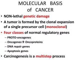
GENES PLAYING ROLE IN CANCER DEVELOPMENT • Oncogenes • Tumor suppressor genes • DNA repair genes
+ - ++ What are the genes responsible for tumorigenic cell growth? Normal Proto-oncogenes Cell growth and proliferation Tumor suppressor genes Cancer Mutated or “activated” oncogenes Malignant transformation Loss or mutation of Tumor suppressor genes
ONCOGENES • Oncogenes are mutated forms of cellular proto-oncogenes. • Proto-oncogenes code for cellular proteins which regulate normal cell growth and differentiation.
Five types of proteins encoded by proto-oncogenes participate in control of cell growth: Class I: Growth Factors Class II: Receptors for Growth Factors and Hormones Class III: Intracellular Signal Transducers Class IV: Nuclear Transcription Factors Class V: Cell-Cycle Control Proteins
Functions of Cellular Proto-Oncogenes 1. Secreted Growth Factors 2. Growth Factor Receptors 4. Nuclear Proteins: Transcription Factors 3. Cytoplasmic Signal Transduction Proteins 5. Cell Growth Genes
A generic signalling pathway
Oncogenes proto-oncogene = ras Oncogene = mutated ras Always activated Always stimulating proliferation
Amino acid substitutions in Ras family proteins (inactivates GTPase) amino acid position Ras gene 12 59 61 Tumor c-ras (H, K, N) Gly Ala Gln normal cells H-ras Gly Ala Leu lung carcinoma Val Ala Gln bladder carcinoma K-ras Cys Ala Gln lung carcinoma Arg Ala Gln lung carcinoma Val Ala Gln colon carcinoma N-ras Gly Ala Lys neuroblastoma Gly Ala Arg lung carcinoma Murine sarcoma virus H-ras ArgThr Gln Harvey strain K-ras SerThr Gln Kirsten strain
Activation mechanisms of proto-oncogenes proto-oncogene --> oncogene
CHROMOSOMAL REARRANGEMENTS OR TRANSLOCATIONS Neoplasm Translocation Proto-oncogene Burkitt lymphoma t(8;14) 80% of cases c-myc1 t(8;22) 15% of cases t(2;8) 5% of cases Chronic myelogenous t(9;22) 90-95% of cases bcr-abl2 leukemia Acute lymphocytic t(9;22) 10-15% of cases bcr-abl2 Leukemia 1c-myc is translocated to the IgG locus, which results in its activated expression 2bcr-abl fusion protein is produced, which results in a constitutively active abl kinase
GENE AMPLIFICATION Oncogene Amplification Source of tumor c-myc ~20-fold leukemia and lung carcinoma N-myc 5-1,000-fold neuroblastoma retinoblastoma L-myc 10-20-fold small-cell lung cancer c-abl ~5-fold chronic myoloid leukemia c-myb 5-10-fold acute myeloid leukemia colon carcinoma c-erbB ~30-fold epidermoid carcinoma K-ras 4-20-fold colon carcinoma 30-60-fold adrenocortical carcinoma
Oncogenes are usually dominant • (gain of function) • cellular proto-oncogenes that have been mutated (and “activated”) • cellular proto-oncogenes that have been captured by retroviruses and have been mutated in the process (and “activated”) • virus-specific genes that behave like cellular proto-oncogenes that have been mutated to oncogenes (i.e., “activated”)
The result: • Overproduction of growth factors • Flooding of the cell with replication signals • Uncontrolled stimulation in the intermediary pathways • Cell growth by elevated levels of transcription factors
Tumor suppressor genes • Normal function - inhibit cell proliferation • Absence/inactivation of inhibitor --> cancer • Both gene copies must be defective
KNUDSON TWO HIT HYPOTHESIS IN FAMILIAL CASES Familial RB (%30) rb RB Normal cells rb RB rb RB LOH Inactivation of a tumor suppressor gene requires two mutations, inherited mutation and somatic mutation. Tumor cells Normal cells
RB RB RB LOH RB Mutation KNUDSON TWO HIT HYPOTHESIS IN SPORADIC CASES Normal Cells RB RB Inactivation of a tumor suppressor gene requires two somatic mutations. Tumor cells
TUMOR SUPPRESSOR GENES Disorders in which gene is affected Gene (locus) Function Familial Sporadic DCC (18q) cell surface unknown colorectal interactions cancer WT1 (11p) transcription Wilm’s tumor lung cancer Rb1 (13q) transcription retinoblastoma small-cell lung carcinoma p53 (17p) transcription Li-Fraumeni breast, colon, syndrome & lung cancer BRCA1(17q) transcriptional breast cancer breast/ovarian tumors BRCA2 (13q) regulator/DNA repair
Daugther cell Gateway Mitosis Growth Factors CELL CYCLE DNA replication Cell cycle inhibitors Control Point CELL CYCLE S
Rb gene • Rb protein controls cell cycle moving past G1 checkpoint • Rb protein binds regulatory transcription factor E2F • E2F required for synthesis of replication enzymes • E2F - Rb bound = no transcription/replication • Growth factor --> Ras pathway --> G1Cdk-cyclin synthesized • Active G1 Cdk-cyclin kinase phosphorylates Rb • Phosphorylated Rb cannot bind E2F --> S phase • Disruption/deletion of Rb gene • Inactivation of Rb protein --> uncontrolled cell proliferation --> cancer
p53 • Phosphyorylated p53 activates transcription of p21 gene • p21 Cdk inhibitor (binds Cdk-cyclin complex --> inhibits kinase activity) • Cell cycle arrested to allow DNA to be repaired • If damage cannot be repaired --> cell death (apoptosis) • Disruption/deletion of p53 gene • Inactivation of p53 protein --> uncorrected DNA damage --> uncontrolled cell proliferation --> cancer
DNA REPAIR GENES These are genes that ensure each strand of genetic information is accurately copied during cell division of the cell cycle. Mutations in DNA repair genes lead to an increase in the frequency of mutations in other genes, such as proto-oncogenes and tumor suppressor genes. i.e. Breast cancer susceptibility genes (BRCA1 and BRCA2) Hereditary non-polyposis colon cancer susceptibility genes (MSH2, MLH1, PMS1, PMS2) have DNA repair functions. Their mutation will cause tumorigenesis.
Molecular mechanisms of DNA double strand break repair BRCA1/2 Van Gent et al, 2001