Download
1 / 63
630 likes | 978 Vues
Evaluation and Management of Primary Amenorrhea. Libby Crockett, MD Department of Obstetrics and Gynecology University of Nebraska Medical Center. Disclosure…. I have no financial conflicts of interest. . Objectives. 1 . Understand the causes of primary amenorrhea

E N D
Evaluation and Management of Primary Amenorrhea Libby Crockett, MD Department of Obstetrics and Gynecology University of Nebraska Medical Center
Disclosure…. • I have no financial conflicts of interest.
Objectives • 1. Understand the causes of primary amenorrhea • 2. Understand how to elicit a pertinent history and perform a focused physical exam to evaluate primary amenorrhea • 3. Understand how to perform and interpret selected diagnostic tests and imaging to evaluate primary amenorrhea
Puberty—The Mechanism • Initiated by release of pulsatile GnRH (hypothalamus) • Specifically see increased pulsatile patterns of FSH & LH (these start during sleep and eventually go throughout the day) • With pulses of GnRH, peaks of estradiol result and eventually menarche appears • By late puberty, the mature HPO axis is intact and ovulation occurs
Timing of Puberty • Major determinant—GENETICS • Also affected by geographic location, exposure to light, general health and nutrition and psychological factors • Recent studies have demonstrated a decline in the age of menarche • Critical Body Weight? • Studies have shown 47.8 kg in general • Shift in body composition to more fat 16-23.5% • Has been linked to protein Leptin
Stages of Pubertal Development • Stages • 1. Accelerated Growth • 2. Breast Development (Thelarche) • 3. Pubarche • 4. Menarche • Generally takes 4.5 years • Differs culturally between Ethnic Groups
Puberty and Ethnicity Wu T, Mendola P, Buck GM. Ethnic differences in the presence of secondary sex characteristics and menarche among US girls: the Third National Health and Nutrition Examination Survey. 1988-1994. Pediatrics. 110: 752, 2002.
Assessing Pubertal Development • Tanner Staging • Developed by James Tanner and originally published in 1968 as an objective way to assess pubertal development. Speroff and Fritz. Abnormal Puberty and Growth Problems. Clinical Gynecological Endocrinology and Infertility: Seventh Edition. 2005. pg 365-392
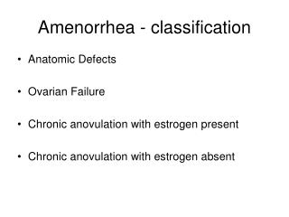
Causes of Primary Amenorrhea • American Society of Reproductive Medicine classifies causes of primary amenorrhea into three distinct groups • Primary Amenorrhea with: • Breast Development (30%) • No breast development AND high FSH (40%) • No breast development AND low FSH. (30%) ASRM Practice Committee. Amenorrhea. FertilSteril 2008.
Causes of Primary Amenorrhea ASRM Practice Committee. Amenorrhea. FertilSteril 2008.
Mullerian Agenesis • Mayer-Rokitansky-Kuster-Hauser (MRKH) syndrome • Complete absence of uterus, cervix and the upper 2/3 of the vagina • Incidence 1/5000 (1/4000-1/10,000 female newborns) • Normal XX Karyotype • Normal ovarian function • Otherwise normal pubertal development • Causes • Mutations in Antimullerian Hormone or Antimullerian Hormone receptor • Association with Wnt gene Deligeoroglou et. Al 2010 & ASRM Practice Committee. Amenorrhea. FertilSteril 2008.
Uterine Development Video • Hill, M.A. (2013) Uterus Development Movie. Retrieved August 5, 2013, from http://php.med.unsw.edu.au/embryology/index.php?title=Uterus_Development_Movie
Mullerian Agenesis • Evaluation: Normal breast development, normal secondary sexual characteristics • Laboratory: Normal XX karyotype, normal LH, FSH • Pelvic Exam: • Normal external genitalia • absence of internal midline structures • + vaginal dimple
Diagnosis Left: MRI showing absence of uterus and vagina
Mullerian Agenesis • Associated Conditions • 1/3 concurrent urinary tract anomalies • Ex: Ectopic kidney, renal agenesis, horseshoe kidney • 12% associated skeletal anomalies • Ex: spinal anomalies, absent digits, webbed fingers, toes • Part of work up needs to include an abdominal CT to evaluate for renal anomalies
Mullerian Agenesis: Treatment • Dilators (Frank and Ingram) • Dilate at a 15 degree angle daily after warm bath for 20 minutes. • Progressively work up to larger dilators • Success defined as non-painful intercourse or vaginal length of 7cm • Studies demonstrate up to an 88% success rate at 19 months of use.
McIndoe Neovagina • Use a skin graft or artificial skin placed over a mold forming a tube with one closed end • Incision made in the vaginal dimple and cavity dissected to level of peritoneum. • Labia majora are sewn together. • Bed rest for 7 days and then mold removed.
Other Forms of Surgical Management • Williams Vaginoplasty • Use labia majora in order to create vagina • Disadvantage—produce shorter vaginal cavity • Sigmoid Vaginoplasty • Pulldown of sigmoid colon to introitis followed by end to end reanastomosis. • Advantages: good vaginal length • Disadvantages: report of foul smelling discharge and odor. • Vecchietti Vaginoplasty • Creation of neovagina by invagination • Small acrylic mold placed in the vaginal dimple. • Abdominal incision made—traction stitches placed on the abdominal peritoneum and attached to the mold. Traction device used to pull mold up 1-1.5 cm per day. Takes approximately 7-9 days.
Transverse Vaginal Septum • Failure of canalization of distal third of the vagina • Most common in upper and middle third of vagina • Diagnosis • Usually present after puberty with amenorrhea and pelvic pain • Can present with hematocolpos, hematometra • Does not bulge with valsalva maneuver • MRI helps with diagnosis
Vaginal Septum Illustration by John Parker. Wheeless, CR. And Roenneburg, M.L. Atlas of Pelvic Surgery http://www.atlasofpelvicsurgery.com/2VaginalandUrethra/6ExcisionofTransverseVaginalSeptum/chap2sec6.html
Imperforate Hymen • Most common obstructive lesion of the female genital tract • 1/1000 female births • Classic appearance of bulging, blue-domed, translucent membrane • Can present with hematocolpos or urinary retention • Differs from vaginal septum in that an imperforate hymen bulges with valsalva • Treatment: Surgical Resection • Hymenectomy versus hymenotomy
Androgen Insensitivity • Incidence 1/60,000 • although 9% of causes of primary amenorrhea • Genetics: X-linked recessive • Phenotype: Female; Genotype: XY • Female external genitalia with small vaginal dimple • Absent uterus and cervix. • Cryptorchidic gonads • Absent axillary and pubic hair • +Breast development • Cause: Mutations in the androgen receptor Deligeoroglou et al 2010
Androgen Insensitivity • Physical Exam • Slim and taller than average female • Large breasts with juvenile nipples • Absent pubic/axillary hair, no acne or other signs of androgen action • May have inguinal hernia • Normal external genitalia, blind vaginal pouch and absence of midline structures • Laboratory: Testosterone in the normal to high male range
Androgen Insensitivity: Removal of Gonads • Location of testicular gonads is variable • Intrabdominal cavity • Labialscotal folds • Inguinal region • Recommend removal after complete pubertal development • Enhance bone maturation and puberty • Recommend at age 16-18 • Once testes removed, treat with hormone replacement therapy • Incidence of neoplasia • 22% incidence of malignancy • Most common histology is Leydig cell hyperplasia
Causes of Primary Amenorrhea HypergonadotrophicHypogonadism Gonadal dysgenesis Current Evaluation of Amenorrhea. Fertility and Sterility 2008 80(3): S219-S225.
Turner’s Syndrome • Classically 45 XO or mosaic • Incidence 2,500-10,000 liveborns • 99% of pregnancies affected end in SAB • Cause: Absence of ovarian determinant genes result in premature loss of germ cells • Fetuses with Turner’s have the same amount of germ cells at midgestation as do 46, XX • As gestation continues, accelerated loss of germ cells occurs • Many XO individuals lose all germ cells prior to birth; less than 15% have enough germ cells to start pubertal process by adolescence
Turner’s Syndrome • Associated Abnormalities • Cardiac Anomalies • Coartation of aorta in 30% of patients, also bicuspid aortic valve, mitral valve prolapse • Recommend echocardiography be performed every 3-5 years • Renal Anomalies • Horseshoe Kidney • Need retroperitoneal ultrasound once diagnosed • Hypothyroidism • 10% of patients with Turner’s Syndrome • Recommend yearly screening of T4/TSH and antibodies • Deafness (audiometry)
Gonadal Dysgenesis: 46XX • Refers to a number of conditions in which abnormal development leads to streak gonads • Incidence: <1/10,000 in women less than 30 • Inherited • Familial inheritance 7-30% • Premutations in the FMR1 gene (Fragile X Syndrome) • 15% of carriers have POF • Associated with autoimmune diseases (18-30%) • Hashimoto’s Thyroiditis, Addison’s disease, hypoparathyroidism, vitiligo • Acquired • Radiation, chemotherapy • Environmental • Childhood viruses
Gonadal Dysgenesis: 46XX • Diagnosis: • >3 months of amenorrhea + FSH in the menopausal range • Ultrasound; >60% of patients have undetectable ovaries by ultrasound. Majority show no follicular growth • DEXA scan in addition to screening for autoimmune diseases • Hormone Replacement • Low-dose estradiol(1/2 mg/day and step up) for 12-18 months before addition of progestogenic agent • Add progesterone in order for regular menstruation. • Fertility • 5-10% of spontaneous pregnancy as patients with gonadal dysgenesiswill cycle inconsistently. • Recommend OCPs in adolescent population to prevent unwanted pregnancy.
Gonadal Dysgenesis: 46 XY Swyer Syndrome • Cause: Associated with mutations in the SRY gene. • Streak gonads present; No testes formation • Therefore Anti-Mullerian hormone and testosterone are not produced thus • Normal uterus and fallopian tubes, female external genitalia • Estrogen also not produced from streak gonads therefore breast development does not occur • Elevated FSH/LH • Streak Gonads need removal as they are at increased risk (25%) for germ cell tumors: most common gonadoblastoma.
Swyer Syndrome Dysgerminomain an adolescent patient with Swyer Syndrome
Causes of Primary Amenorrhea HypogonadotropicHypogonadism Current Evaluation of Amenorrhea. Fertility and Sterility 2008 80(3): S219-S225
Prolactinoma • Most common cause of pituitary related amenorrhea • Mechanism • Elevated PRL levels can suppress hypothalamic GnRH secretion • Higher the PRL level, the greater disruption of the menstrual cycle • Rule out hypothyroidism! • Medications? • Imaging • MRI of pituitary fossa if PRL is >100ng/mL OR if visual symptoms • Treatment • Bromocriptine/Cabergoline • 80-90% of hyperprolactinemia will resolve and 80% of microadenomas will shrink • Resort to transsphenoidal surgery if medical therapy fails.
Prolactinoma Fig. 2. a MRI: microadenoma 8 mm (arrow) in a 14-year-old female. b MRI: macroadenoma in a 15-year-old male (pretreatment). c MRI: empty sella post-treatment in the same patient
Other Pituitary Causes of Amenorrhea Deligeoroglou et al. Evaluation and management of adolescent amenorrhea. Annals of the New York Academy of Sciences. 2010; pg 23-32
Hypothalamic Amenorrhea • Functional hypothalamic amenorrhea • Stress, nutrition, and exercise related • Alterations in normal pulsatile release of GnRH. • Mechanism • Complex interplay between neuropeptides • Leptin • Lower levels of leptin (malnourished, anorexia) seem to decrease amount of leptin and cause amenorrhea. • Cortisol • Stress related levels CRH interfere and inhibit GnRH • Exercise • Chronic imbalance between calorie intake and consumption lead to hypothalamic dysfunction
Hypothalamic Amenorrhea • Primary Amenorrhea and Eating Disorders • Despite treatment, adolescents with eating disorders and primary amenorrhea progress through puberty at a slowed rate • Estimate weight at which menarche will resume by prepubertal weight
Female Athlete Triad • Established as a diagnosis in 1992 • Amenorrhea, osteoporosis, and eating disorder among female athletes • Most common: gymnastics, ballet, and long-distance running • Bone Mineral Density evaluation • Athletes general have higher BMD • A Z-score less than -1.0 requires evaluation
Female Athlete Triad • Counseling about Eating Disorders • Screening tests: • Eating Attitudes Test • SCOFF questionnaires • Dietician • Minimal goal of 30 kcal/kg of lean body mass • Dairy, iron, and protein rich foods • Referral to psychologist • Role of Oral Contraceptives • Some improvement in BMD but does not restore bone mass to age –matched controls • Need to address underlying pathology—focused counseling with regard to nutrition and psychology
Kallman Syndrome • Cause • X-linked recessive mutation in the KAL gene • Codes of an adhesion moleculeresults in lack of migration of GnRH neurons from the olfactory placcode. • Characteristics • Hypogonaotrophic hypogonadism • Anosmia • Midline facial defects • Occasional renal agenesis • See absence of pubertal development and primary amenorrhea • Treatment • Hormone replacement therapy to promote sexual maturation • Fertility is possible using IM gonadotropins
Polycystic Ovarian Syndrome • 5-10% of adult women and increasing in prevalence in the adolescent population • Diagnosis • At least 2 of the following: • Chronic Anovulation • Clinical or biochemical evidence of excess androgen • Polycystic ovaries on ultrasound • Typically present with secondary amenorrhea/oligomenorrhea but represent 3% of diagnoses of primary amenorrhea • Important to diagnose given metabolic abnormalities • Recent study of adolescent population showed 62% had already developed insulin resistance • Dyslipidemia • Obesity