Download
1 / 75
981 likes | 2.56k Vues
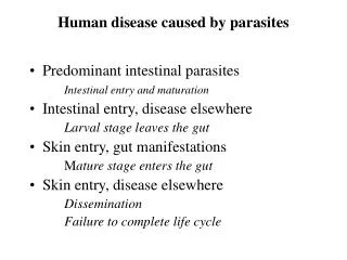
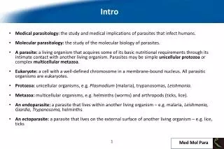
Medical Helminthology : Flatworms—human parasites. Lecturer: ass. prof. Tetyana Bihunyak. According to the way of development helminths are classificated into biohelminthes and geohelminthes .

E N D
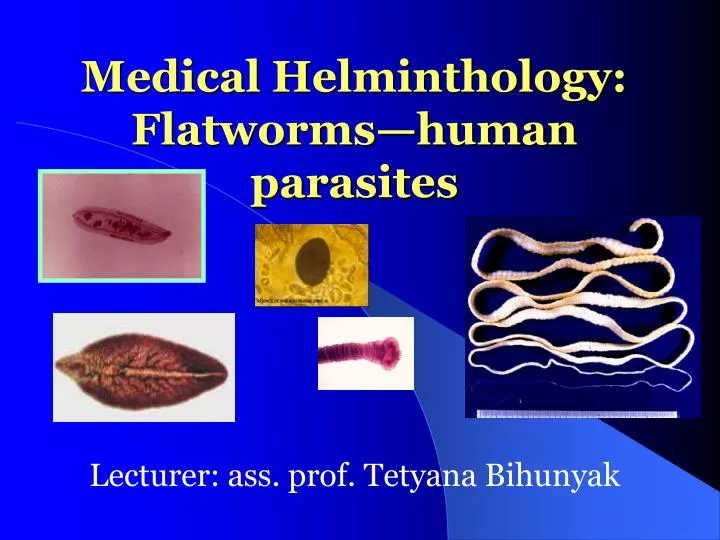
Medical Helminthology:Flatworms—human parasites Lecturer: ass. prof. Tetyana Bihunyak
According to the way of development helminths are classificated into biohelminthes and geohelminthes. • Geohelminthes develop without intermediate host. Soil is the best environment for their egg's development. Humans are infected through dirty fruits and vegetables, which contain geohelminthe's eggs (Ascaris lumbricoideus). • Biohelminthes have complete life cycle with definitive and intermediate hosts. There are trophycal connections between definitive and intermediate hosts (for example, Taenia solium).
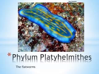
General characteristic of Flatworms (Phylum Plathelminthes) • The flatworms consists of 12, 200 species, including classes of parasitic worms: Trematoda, Cestoda • All flatworms are acoelomate, triploblastic, bilaterally symmetrical; flattened dorsoventrally • They have a definite head at the anterior end • Their bodies are solid: the only internal space consists of the digestive cavity. They have no anus; a single opening to the digestive system serves as both mouth and anus • Wastes probably move out of flatworms mostly by diffusing across the general body surface • The most of flatworm species are hermaphrodites.
General characteristic of Class Trematoda 1) Flattened dorsoventrally (leaf-like). 2) Unsegmented. 3) Body is covered by cuticle. 4) Organs of fixation: oral sucker, ventral sucker. 5) Organs and systems: digestive system, excretory system, nervous system. Genital system: Trematodes are hermaphrodites except genus Schistosoma. 6) The life cycle is passed in two hosts (alternation of hosts) and has sexual and asexual stages.
Schistosomiasis is second only to malaria in human impact among tropical diseases and is the third most prevalent parasitic disease in the world.
Schistosomiasis/Distribution • Schistosoma haematobium, S. mansoni infections: in sub-Saharan Africa • S. mansoni remains endemic in parts of Brazil, Venezuela and the Caribbean • S. japonicum still occurs in China, Indonesia & the Philippines
BLOOD FLUKES - genus SCHISTOSOMA • Schistosoma mansoni andSchistosoma japonicum cause Hepatosplenic Schistosomiasis. • Schistosoma haematobium causes Urinary Schistosomiasis. • Localization: venous vessels of bowel, liver, and bladder. • Morphology: atypical trematodes which the adult female nesting within a specialized groove in the body of the larger male.
Schistosome adult worms • Male/female pair copulate throughout life-produce eggs • Females resides in canal-Important for maturation • Some differences among species • Worm pairs can live for more than 10 years in a host • Pair migrate back against the blood flow to the mesenteries around the intestine.
BLOOD FLUKES • Infective stage for human: cercariae. • Definitive host:man. • Intermediate host:snail. • Mode of transmission:penetration of skin by cercarie.
Cercaria in the water • It is composed of a body 125 m long by 25 m in diameter to which a 200 m long tail is attached. • First escape into the hemolynph and then through the snail’s integument • Swim into the surrounding water to find their definitive host • Swims by alternating side-to-side rhythmic contractions
How do you get schistosomiasis ??? How about total body immersion …. In a canal off the Nile, just southwest of Cairo, Egypt In a storage reservoir just outside of Belo Horizonte, Brazil
In endemic areas, most at risk are school-age children, women, and those involved in occupations such as irrigation, farming and fishing.
Then there is the North American schistosome-induced pathology --- Duck schistosomes Raccoon schisto;… etc. Sign on the door leading out of a resort to Lake Bemidji, in Minnesota (the advice can’t hurt, but may not help either Cercarial Dermatitis
The 2 faces of schistosomiasis ‘Intestinal’ asymptomatic schistosomiasis at the Egyptian village level Egyptian boy with hepatosplenomegaly, ascites fluid build-up and superficial collateral circulation (NAMRU-3 clinical ward in Cairo)
BLOOD FLUKES • Clinical manifestationsof Hepatosplenic Shistosomiasis: eosinophilia, polyps in colon, fever, anorexia, weight loss, anemia, portal hypertension; cirrhosis of liver; pruritic skin rash. Eggs go back through portal circulation to liver, causing hepatomegaly, liver tenderness. • Clinical manifestationsof Urinary Schistosomiasis: eosinophilia, hematuria, terminal dysuria (pain, difficulty at the end of urination); obstructed urine flow.
The abdomen of an 11-year-old boy with intestinal schistosomiasis with the size and extent of the liver and spleen marked, indicating the severity of infection. The disease has caused a stunting of the boy's growth, he is only 120cms tall and weighs 22 kg.
BLOOD FLUKES • Laboratory diagnosticsof Hepatosplenic Schistosomiasis: eggs with lateral spine in feces • Laboratory diagnostics of Urinary Schistosomiasis: eggs with terminal spine in urine • Prevention:involves proper disposal of human waste anderadication of the snail host when possible. Swimming in endemic areas should be avoided.
S. manosni egg: prominent lateral spine Ovoid (140X61µ) S. haematobium egg: prominent terminal spine, ovoid (150X62µ) S. japonicum egg: lateral spine obscured, round (100X60µ) Morphology of the Eggs of the 3 Key Schistosomes That Infect Humans
Control programs • Large scale population based chemotherapy • Environmental modification • Controlling snail habitat • Use of molluscicides • Behavioral modification • Difficult and costly to sustain
How can I prevent schistosomiasis? • Avoid swimming or wading in fresh water when you are in countries in which schistosomiasis occurs. • Swimming in the ocean and in chlorinated swimming pools is generally thought to be safe.
LUNG FLUKE: PARAGONIMUS WESTERMANI - an agent of paragonimiasis • Distribution: Far East, Central America, Africa, and India. • Morphology: an egg-like form of the body, from 7,5 to 16 mm.
LUNG FLUKE • Mode of transmission: ingestion of metacercaria in crabs or crayfish. • Final hosts: carnivoirous mammals, pigs, humans. • Intermediate hosts: 1) snail (sporocyst, redia, cercaria); 2) crabs or crayfish (metacercaria). • Infective stage: metacercariae
LUNG FLUKE • Clinical disease: a chronic cough with bloody sputum, dyspnoa, pleuritic chest pain, and pneumonia. • Laboratory diagnosis: eggs in sputum or feces. • Prevention: cooking crabs and crayfish properly.
BILIARY (LIVER) FLUKES • CLONORCHIS SINENSIS–oriental small biliary (liver) fluke, causes Clonorchiasis. • Distribution:endemic in Far East, China, Japan, and Vietnam. • Localization:bile ducts, gallbladder, and pancreas.
CLONORCHIS SINENSIS Morphology: the adult worms are 1 to 2 cm; the eggs are small, brownish.
CLONORCHIS SINENSIS • Transmission:fecal-oral (ingestion of contaminated raw, frozen, dried, pickled, and salted fish). • Infective stage:metacercariae. • Final hosts:carnivorous mammals and humans. • Intermediate hosts: • 1- snail (miracidium, sporocyst, rediae, cercariae), • 2 - fish Cyprinidae genus- the family that includes carp and goldfish (metacercariae).
CLONORCHIS SINENSIS • Clinical disease:cholecystitis and cholelithiasis, hepatic colic, associated with profound weight loss and diarrhea. An individual fluke may live for 15-30 years in the liver. In humans a heavy infestation of liver flukes may cause cirrhosis of the liver and death. • Laboratory diagnosis:immature eggs in feces • Prevention:adequate cooking of fish and proper disposal of human waste
FASCIOLA HEPATICA • an agent of fascioliasis. Distribution: endemic in Far East. • Localization: bile ducts, gallbladder, and pancreas. • Morphology: large size (3-5 cm) and conical form of the body; possess sucking disks (oral and abdominal) that provide them motion. Multibranched uterus is situated under the abdominal sucking disk. Testis are branched too and situated in the middle part of the body.
FASCIOLA HEPATICA Life-cycle: • Final host - herbivorous mammals (horses) and humans. • Intermediate host — the snail Limnea truncatula. • Transmission:fecal-oral (ingestion of water, some non-water plants and vegetables, which contain adolescariae). • Invasive stage:adolescariae.
FASCIOLA HEPATICA • Clinical disease:Parasites obstruct bile ducts and lay eggs within them, leading to cholelithiasis (gallstones). Biliary obstruction can occur, sometimes causing biliary cirrhosis. • Diagnosis: immature eggs in feces. An egg has large sizes, thin membrane, yellow color and small cover in one pole. • Prevention: involves not eating wild aquatic vegetables.
OPISTHORCHIS FELINEUS • small biliary fluke, causing Opisthorchiasis. • Distribution: Siberia. • Morphology: flat, the length of the body 4-13 mm. In the middle part of the body there is a branched uterus. Behind it there is a round ovary. There is a roseolla-like testis in the back of the uterus - a diagnostic sign of this worm.
OPISTHORCHIS FELINEUS • Life-cycle:Final host - carnivorous mammals and humans. • Intermediate hosts: 1) snail Bithynia leachi genus; 2) fish. • Transmission: ingestion of contaminated raw, frozen, dried, pickled, and salted fish, which contains metacercariae.Invasive stage: metacercariae in fish muscles. • Localization: bile ducts, gallbladder, liver.
Clinical disease: cholecystitis and cholelithiasis, hepatic colic, cirhosis. Clinical picture is very similar to Clonorhis infection. Infection can lay dormant for several years before presenting clinically. • Diagnosis: immature eggs in feces, in fluid from biliary drainage, or duodenal aspirate. Eggs are 15-30 mcm in sizes, have oval form and yellow color. The outer membrane is thick, and there is a cover in the front of the egg. The internal structure of the egg is microgranular. • Preventioninvolves not eating undercooked or contaminated raw, frozen, dried, pickled, and salted fish; eradication of snail hosts when possible.
DICROCOELIUM LANCEATUM – causes Dicrocoeliasis • Distribution: worldwide. • Localization: bile ducts, gallbladder and liver of mammals (cattle, horses). Very rare in humans. • Morphology: the worms are 1 cm long with lanceolate form of the body; the intestine (gut) has two nonbranched channels which are situated in the lateral sides of the body. Two round testis are situated in the front of the body - the diagnostic sign of this worm.
DICROCOELIUM LANCEATUM Life-cycle: • Final host - herbivorous mammals (cattle, horses). • Intermediate hosts: 1) the snail of Zebrina and Helicela genus, 2) ants Fornica genus. • Transmission: ingestion of plants with the ants, which contain metacercariae. • Diagnosis: immature eggs in feces. An egg have oval form, smooth membrane, brown color, a cover is present in the front end. • Prophylactics: eradication of the snails, ants when possible; dehelmithization of cattle.
Tapeworms (Cestoda) • consist of a rounded head, called a scolex, and long strobila or chain of proglottids (multiple segments) of varying stages of maturity. • They have no digestive tract of its own at any point in its life cycle. • The scolex has specialized means of attaching to the intestinal wall, namely suckers, hooks, or sucking grooves. • All cestodes have stage of larva and stage of oncosphere in the life cycle.
General Body Shape of a Tapeworm • Scolex • Neck • Strobila made up of proglottids
Scolices Suckers and hooks
Scolex (pl: Scolices) • Bothria – with slit-like groove with weak suction powers and usually two in number
Neck • Undifferentiated stem cells that give rise to proglottids in strobila.
Proglottids • Unique structure of Cestodes • Contains both male and female organs • Essentially a whole reproductive package in one segment of the strobila.
Integument • Microtriches (like Microvilli of vertebrate small intestine), on surface of proglottid. • Absorb nutrients
Proglottid RED =Male BLUE = Female
Systematics • Order Pseudophyllidea • Family Diphyllobothriidae • Diphyllobothrium latum • Order Cyclophyllidea • Family Taeniidae • Taenia saginata, T. solium, T. multiceps, T. hydatigena, T. pisiformis, T. taeniaeformis, T. ovis • Echinococcus granulosus • Family Dilepididae • Dipylidium caninum • Moniezia benedeni, M. expansa • Anoplocephala spp. • Order Cyclophyllidea • Family Taeniidae • Taenia saginata, T. solium, T. multiceps, T. hydatigena, T. pisiformis, T. taeniaeformis, T. ovis • Echinococcus granulosus • Family Dilepididae • Dipylidium caninum • Moniezia benedeni, M. expansa • Anoplocephala spp.
Definitive Host Adult Life Cycles Egg } Coracidium Intermediate Host Oncosphere Cysticercus Hydatid Cysts
Taenia solium • The adult form of T. soliumcauses taeniasis solium.T. solium larvae cause cysticercosis. • Distribution Teniasis and cysticercosis occur worldwide but is endemic in areas of Asia, South America, and eastern Europe • MorphologyT. solium can be indentified by its scolex with 4 suckers and circle of hooks and by its gravid proglottids, which have 7-12 primary uterine branches. Larva of T.solium called cysticercus. A cysticercus consist of a pea-sized fluid-filled bladder with an invaginated scolex.
Taenia solium Life cycle • Transmittion:fecal-oral • Definitive hosts – humans • Intermediate hosts - pigs • Humans can be infected by eating raw or undercooked pork containing the larvae cysticercus. • In the small intestine, the larvae attach to the gut wall and take about 3 months to grow into adult worm. • The gravid terminal proglottids detach daily, are passed in the feces.