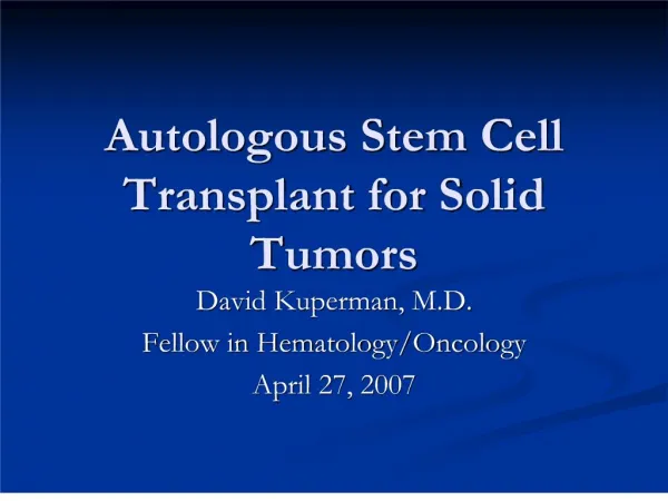
PEDIATRIC SOLID TUMORS
PEDIATRIC SOLID TUMORS. Prof. Dr. Rejin KEBUDİ I.U., Cerrahpaşa M. F., Dept. Of Pediatrics & I.U., Oncology Institute, Div.Pediatric Hematology-Oncology. 65.8%. Survival (%). Time (Months).

PEDIATRIC SOLID TUMORS
E N D
Presentation Transcript
PEDIATRIC SOLID TUMORS Prof. Dr. Rejin KEBUDİ I.U., Cerrahpaşa M. F., Dept. Of Pediatrics & I.U., Oncology Institute, Div.Pediatric Hematology-Oncology
65.8% Survival (%) Time (Months) Turkish Pediatric Oncology Group (TPOG) & Turkish Pediatric Hematolgy Society (TPHD) Pediatric Tumor Registry, 2002-2008 Survival Rate (7 Year) T.Kutluk & A. Yesilipek, on behalf of TPOG/TPHD
Survival (%) Time (Months) Turkish Pediatric Oncology Group (TPOG) & Turkish Pediatric Hematolgy Society (TPHD) Pediatric Tumor Registry, 2002-2008 Survival rates in different tumor types T.Kutluk & A. Yesilipek, on behalf of TPOG/TPHD
Cancer In Children Rhabdomyosarcoma Non-RMS STS
Turkish Pediatric Oncology Group (TPOG) & Turkish Pediatric Hematology Society (TPHD) Pediatric Tumor Registry, 2002-2008 Tumor Type n % I Leukaemia 2614 31,3 II Lymphomas and Reticuloendothelial Neoplasm 1552 18,6 III CNS and Miscellaneous Intracranial and Intraspinal Neoplasm 1084 13,0 IV Sympathetic Nervous System Tumors 622 7,4 IX Soft-TIissue Sarcomas 505 6,0 V Retinoblastoma 193 2,3 VI Renal Tumors 470 5,6 VII Hepatic Tumors 122 1,5 VIII Malignant Bone Tumors 509 6,1 X Germ Cell, Trophoblastic and Other Gonadal Neoplasm 371 4,4 226 2,7 XI Carcinomas and Other Malignant Epithelial Neoplasm 87 1,0 XII Other and Unspecified Malignant Neoplasm Total 8355 100,00 T.Kutluk & A. Yesilipek, on behalf of TPOG/TPHD
Warning Signs of Cancer in Children Continued, unexplained weight loss Headaches with vomiting in the morning Increased swelling or persistent pain in bones or joints Lump or mass in abdomen, neck, or elsewhere Development of whitish appearance in pupil Recurrent fevers not caused by infections Excessive bruising or bleeding Noticeable paleness or prolonged tiredness
Çocuk kanserlerinde başlıca belirti ve bulgular • Solukluk, halsizlik • Sık ateşlenme • Kilo kaybı • Gelişme geriliği • Kanama bulguları (peteşi, ekimoz) • Burun, dişeti kanamaları
Çocuk kanserlerinde başlıca belirti ve bulgular • Başağrısı, kusma • Ateşsiz havale geçirme • Dengesizlik, yürüme bozukluğu • Gözde kayma, görme bozukluğu • Göz bebeğinde parlaklık
ÇOCUKLUK ÇAĞI TÜMÖRLERİNDE UYARICI BULGU VE BELİRTİLER • Kemik, eklem ağrıları • Enfeksiyon tedavisine rağmen sebat eden öksürük, nefes darlığı • İdrarda kan gelmesi • İdrar ve dışkılamada zorlanma
Yorgunluk Solukluk Ateş Kanama bulguları Ağırlık kaybı Geçmeyen öksürük Tedaviye rağmen geçmeyen enfeksiyon bulguları Lenfadenomegali Kafa içi kitleler Mediasten kitleleri Abdominal kitleler Urogenital kitleler Ekstremite kitleleri Endokrin belirtiler Diğer belirtiler Çocuk kanserlerinde başlıca belirti ve bulgular
TANI • Muayene • Kan tetkikleri (TKS, förmül, biyo.,tm belirteçleri) • Kemik iliği tetkiki (bazı kanserlerde) • Görüntüleme yöntemleri • Patolojik tanı için örnek alınması
PEDIATRIC ÇAĞI BRAIN TUMORS • Mostly • embriyonal • infratentorial (≤2 yrsupratentorial) • CNS seeding • Treatment • Surgery Radiotherapy Chemotherapy
-Supratentorial Astrositomlar (yüksek grad %10,düşük grad %20) SPNET Ependimom -Infratentorial Medulloblastom Beyin sapı gliomu Epandimoma Serebellarastrositoma -Midline Optik kiazmagliomları Kraniofarinjioma Pineal bölge tümörleri
SUPRATENTORİAL HEMİSFERİK • Konvulziyon • Hemiparezi • Bir yanda duyu kusuru • Hiperrefleksi • Frontal lob-kişilik ve motor fonksiyonlar • Parietal lob-okuma, kontralateral ekstremiteler • Temporal lob-afazi, parsiyel kompleks konv. • Oksipital lob-görme alanı bozukluğu
İNFRATENTORİAL(arka çukur, beyin sapı, serebellar hemisfer) • Ataksi • Dismetri, disdiadokinezi • Beyin sapı-kranial sinir tutulumları hemiparezi • Serebellar hemisferlerde-lateralizasyon bulgusu (appendiküler dismetri)
Medulloblastoma Evreleme Kranium MR Spinal aks MR kontrastlı BOS sitoloji
LOKALİZE BULGU VE BELİRTİLERSUPRATENTORİAL ORTA HAT • Optik kiazma tm-görme alanı defekti • Hipotalamus (SÇ) diensefalik sendrom • Kraniofarenjioma-endokrin sorunlar görme alanı boz hidrosefali • Pineal bölge tm- Parinaud sendromu yukarı bakış kısıtlı dilate pupiller konverjans nistagmusu kapak retraksiyonu
Kebudi R,Upadhyaya M,Tuncer S,et al. A novel mutation in the NF-1 gene in two siblings with neurofibromatosis type 1 and bilateral optic pathway glioma. Pediatric Blood and Cancer 2008 OPTİC GLİOMA NF EUROPE 2012 İstanbul
GCT GLİOM
80 60 40 20 0 SEER Data - 5-year relative survival rate by histology and time period 1975-85 74 1985-95 70 65 57 60 55 56 50 47 39 Astrocytoma PNET Other gliomas Ependymoma All CNS
PEDIATRIC LYMPHOMAS%60 Non-Hodgkin , % 40 Hodgkin Hodgkin Lymphoma Reed Sternberg hücresi
Non-Hodgkin lymphomas- fastest doubling time (Burkitt)- tümör lysis syndrome!
Non-Hodgkin : nonlenfoblastik B cell Burkitt/Burkitt Treatment: Chemotherapy
Neuroblastoma • Treatment • Surgery • Chemot.+ Radiotherapy • Biologic tr. • Retinoik asid • Fenretidine • GD + Il-2
Nöroblastoma SonuçlarımızKebudi R, et al. Istanbul Pediatric Oncology Group Neuroblastoma results. 40th Congress of the International Society of Pediatric Oncology, Berlin, 2008. SAĞKALIM 5 ve 10 yıl S Tüm grup %73 ve 54 10 yıl survival Evre I,II % 100 Evre III % 54 Evre IV % 26 (p= 0.0001 ). Yaş <12 ay < 18 ay • 173 hasta, median yaş 26 ay (1-186). Primer Yerleşim • abdomen 125 (surrenal 110) (73%) • mediastinum 22 (12.2%) • paraspinal 18 (10%) • Diğer 8 (4.8%) • Evre: (INSS) • Evre I 16 (9%) • Evre II 20 (12%) • Evre III 32 (19%) • Evre IV 101 (58 %) • Evre IVS 4 (2%).
-Wilmstm Treatment Surgery radikal nefrektomi Chemot. Radioth. (stage3,4;stage 1,2 poor histology)
LIVER TM • Hepatoblastoma • HCC • FNH • Hamartom • İnf. Myofibroblastik tm
Hepatoblastoma - SIOPEL Experience SIOPEL TREATMENT STRATEGY B I O P S Y Pre-operative chemotherapy Post-operative chemotherapy DELAYED SURGERY 2 months 2 - 3 months
SIOPEL 2pilot study -Treatment results - 5-year survival data by risk category 1.0 CDDP alone 0.8 Standard risk 0.6 % 0.4 High risk 0.2 0.0 6 12 18 24 30 36 42 48 months Overall survival Progression free survival
Intraabdominal malignancies • Nöroblastom • Wilms • Lenfoma • Rabdomyosarkom • Ekstraskeletal Ewing • GCT vd. • EVALUATION • Ultrason • Direkt grafiler • BT / MRI • Hemogram • Kemik iliği • Biyokimya • Tümör belirteçleri:NSE,VMA, AFP,b-HCG • İğne/trucut biyopsileri • Laparaskopi/Laparatomi
SOFT TISSUE SARCOMAS Allages % 0.7 2/105 < 15 yr: Allmalignancies % 7 8.8/106 NonRMS % 3 RabdomiyosarkomMedianage 5yr--(2-6) ve (15-19) NONRabdomiyosarkoma Ewingsarcomafamilytm Other
RABDOMIOSARCOMA 1. >% 50 Rabdomiyosarkom 2.Survival % 25( 1970) % 70 (2000) Tedavi:S ± RT + Chemo 3. Prognostic factors
Rabdomiyosarkom - Yerleşim Head and neck Paramengeal Orbital NPNOBB GenitourinaryParatestiküler Vaginal, uterus Mesane/prostat (MP) Extremities Other
İ.Ü.Onkoloji Enstitüsü, Pediatrik Onkoloji B.D.1989 –2002 Demografik veriler N=95, E:K 1:1, Median yaş 5 (1-18) yaş 7 yıllık Sağkalım % 61 Evre Sıklık Sağkalım(7 y)Histoloji Sıklık S (7y) Evre 1 % 29 %100 Alveolar /ind % 37 %49 Evre 2 % 15 %66 Embryonal %46.5 %70 Evre 3 % 48 %62 Botrioid %16.5 %46 Evre 4 % 8 %0 Yerleşimorbita,NPBB % 100, Paratestiküler%90, vagen-uterus % 80, Mesane-prostat % 51, paramenengeal %42,ekstremite %27 Kebudi R,et al.Evaluation of children with rhabdomyosarcoma. Proceedings of American Society of Clinical Oncology 22: 802, 2003
OSTEOSARCOMA • Most 10-20 yr • metaphysis • distal femur, proximal tibia ve proximal humerusta • % 20metastaz at diagnosis (lungs, bone) • Treatment Neoadjuvan chemo+S + Adjuvan chemo
Langerhans Cell Histiocytosis Kebudi,R., Ayan,İ., Görgün,Ö,Özger,H.,Darendeliler E:Evaluation of 36 children with Langerhans cell histiocytosis with bone involvement. Medical and Pediatric Oncology 41: 338, 2003
LCH AML Tuncer S, Kebudi R, Peksayar G, et al. Congenital Mesenchymal chondrosarcoma of the orbit:Ophtalmology 111:1016-22, 2004
Mediastinal masses Anterior Timus Tiroid Teratom (Germ hücreli tümörler) Lenfoma (Hodgkin,NHL) T cell leukemia Middle(%30) Lenfomalar (Hodgkin,NHL) Posterior Mediastinum (%40) Nöroblastom, Nörofibrom/Schwannom