Download

1 / 22
220 likes | 400 Vues
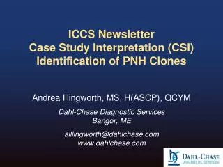
ICCS e-Newsletter CSI Spring 2014. Hong Lin, PhD and Cristina McLaughlin, MD APP-Unipath Denver, Colorado. History and Physical Examination 28-year old African American female with painful lymphadenopathy Palpable lymph nodes in the right groin. Specimens Submitted for Analysis

E N D
ICCS e-Newsletter CSISpring 2014 Hong Lin, PhD and Cristina McLaughlin, MD APP-Unipath Denver, Colorado
History and Physical Examination 28-year old African American female with painful lymphadenopathy Palpable lymph nodes in the right groin Specimens Submitted for Analysis • Peripheral blood received in EDTA. • One tube of bone marrow aspirate received in NaHep for Flow Cytometry testing • One tube of bone marrow aspirate received in EDTA for morphologic examination. • Trephine needle core biopsies of bone marrow (2.4 cm in length by 0.2 cm in diameter) received in B+ for fixation and decalcification. • Right groin lymph node received in RPMI without fixative for flow cytometry testing, as well as portions in formalin for morphologic examination.
WBC Automatic Differential No atypical cells observed on smear evaluation by hematopathologist.
Flow Cytometric Studies Flow cytometric immunophenotyping was performed on right groin lymph node sample and bone marrow aspirate received in NaHep, to “rule out lymphoma”. Data acquisition was performed on a 10-Color Gallios Flow cytometer from Beckman Coulter. Flow cytometric data was analyzed with Kaluza software from Beckman Coulter, using two standard screening panels (Tube 1 and 2), with two add-on custom tubes.
Flow Cytometric Data Analysis – Tube 1 Specimen: right groin lymph node Maroon = abnormal lymphocytes Orange = polyclonal B lymphocytes Blue = polytypic T lymphocytes Non-B lymphocytes bleed into the monocytic area, with high side scatter and bright CD45 Polyclonal B lymphocytes B lymphocytes B lymphocytes Non-B lymphocytes Non-B lymphocytes The CD45/side scatter plot shows mostly lymphocytes, many which show high side-scatter, causing bleed into the monocytic gate. The B lymphocytes (orange) are clearly polyclonal, with a kappa:lambda ratio = 1.8. The non-B lymphocytes do not exhibit CD10 expression.
Flow Cytometric Data Analysis – Tube 2 Maroon = abnormal lymphocytes Orange = polyclonal B lymphocytes Blue = polytypic T lymphocytes Specimen: right groin lymph node Mixture of polytypic T lymphocytes and atypical lymphocytes Unremarkable T lymphocytes Atypical T lymphocytes The remainder of lymphocytes consist of two populations of non-B lymphocytes. There is an unremarkable mixture of polytypic T lymphocytes (blue population), with a mixture of CD4 and CD8 positivity. There is also an atypical cell lymphocytic population (maroon) that expresses higher side scatter and CD4, but is negative for surface CD3 and CD8.
Flow Cytometric Data Analysis – Tube 2 Specimen: right groin lymph node Maroon = abnormal lymphocytes Orange = polyclonal B lymphocytes Blue = polytypic T lymphocytes The atypical lymphocytes (maroon) are negative for CD5, CD7, CD34, and CD56, but brightly positive (as compared to normal T lymphocytes) for CD2. Because of the atypical lymphocytic population, two additional add-on tubes to analyze for T cell markers and for CD30 were added.
Flow Cytometric Data Analysis – Tube 3 Specimen: right groin lymph node Maroon = abnormal lymphocytes Orange = polyclonal B lymphocytes Blue = polytypic T lymphocytes The atypical lymphocytes (maroon) are negative for TCRαβand TCRγδ, as well as surface CD3, while showing bright CD2 as compared to the normal T lymphocytes, which are predominantly TCRαβpositive.
Flow Cytometric Data Analysis – Tube 4 Specimen: right groin lymph node Maroon = abnormal lymphocytes Orange = polyclonal B lymphocytes Blue = polytypic T lymphocytes The atypical lymphocytes (maroon) show variable positivity for CD30, CD25 and CD52.
Key Immunophenotypic Findings • The atypical cells show the following findings: • Cells are negative for pan B cell markers. • Cells express CD4, CD2. • Cells are negative for all other T cell markers, such as CD5, CD7 and surface CD3. • There is no expression of TCRα/β and TCRγ/δ • CD30, CD25, and CD52 expression are heterogeneous.
Lymph Node Morphology Findings • Microscopic Description: • Large, anaplastic lymphoid cells • Eosinophilic cytoplasm • Large irregular nuclei with single or multiple macronucleoli • Rare Hallmark cells • Multiple foci of necrosis • Immunohistochemical Stains: • CD30 is diffusely positive in the large neoplastic lymphocytes • ALK1 is diffusely positive in the large neoplastic lymphocytes
ALK-1 immunostain CD30 immunostain
Bone Marrow Findings • Bone Marrow Morphology: • Trilineage hematopoiesis with appropriate maturation. • Core biopsy with 50% cellularity; mildly hypercellular for age. • Myeloid to erythroid ratio is normal at 3.5:1. • Blasts are not increased; no lymphoid or plasma cell aggregates. • Megakaryocytes are normal in number and morphology. • Immunohistochemistry analysis: • No significant CD30 or ALK-1 positivity observed. • Flow Cytometry studies on bone marrow aspirate: • No Flow Cytometric evidence of hematolymphoid neoplasm. Green = granulocytes Purple = monocytes Blue = lymphocytes Orange = hematogones Red = blasts Gray = debris
Diagnosis: Anaplastic Large Cell Lymphoma (ALCL), ALK positive • In this particular case, though absolute final classification could not be made on flow analysis, the identification of an abnormal T cell population with CD30 expression helped narrow the immunostain workup and accelerated diagnosis by greater than 24 hours. Identification of circulating ALCL cells by flow analysis can also help in identifying patients at high risk of relapse, similar to minimal residual disease studies in leukemia patients, and can help triage patients biopsied by fine needle aspiration. • By flow analysis, if the cells aren’t too large to survive processing, ALCL tumor cells will be positive for CD30 and bright CD45, with many cells bleeding into the monocyte gate on the CD45/SS plot, as we see in our analysis. Given this tendency, an analysis strategy that examines ungated populations as well as gated may yield the greatest sensitivity.
Anaplastic Large Cell Lymphoma (ALCL), ALK positive • WHO 2008 Classification Definition: • T cell lymphoma • Exhibits a translocation involving the ALK gene, with expression of ALK protein and CD30. • Epidemiology: • 3% of adult non-Hodgkin lymphomas • 10-20% of childhood lymphomas • Most frequent in first three decades of life, with a male predominance (M:F ratio 1.5:1)
Anaplastic Large Cell Lymphoma (ALCL), ALK positive • Clinical Features • Patients typically present with advanced stage disease, with peripheral and/or abdominal lymphadenopathy • Often extranodal infiltrates, including skin, bone, soft tissues, lung and liver • High fever • Bone marrow involvement ranges from 10-30% • Prognosis: • The specific translocation does not appear to yield prognostic significance. • ALCL, ALK+ has a favorable prognosis compared with ALCL, ALK negative, with a 5 year survival approaching 80%.
Anaplastic Large Cell Lymphoma (ALCL), ALK positive • Morphology • Cell size varies from small to very large • Eccentric, horseshoe- or kidney-shaped nuclei • Eosinophilic region near the nucleus • Nuclear chromatin is clumped or dispersed with basophilic nucleoli. • Basophilic or eosinophilic cytoplasm • Tumor cells often grow within sinuses and can resemble metastatic tumor. • Small cell pattern also recognized (represents 5-10% of ALCL, ALK+) • Molecular /Cytogenetic • TCR gene rearrangement • Chromosomal abnormalities: • t(2;5)(p23;q35)—NPM-ALK (84%)(IHC shows nuclear and cytoplasmic positivity) • t(1;2)(q25;p23)---TPM3-ALK (13%)(IHC shows diffuse cytoplasmic positivity with peripheral intensification) • Other variant translocations
Anaplastic Large Cell Lymphoma (ALCL), ALK positive • Immunophenotype • CD3 negative in more than 75% of cases • CD2, CD5 and CD4 positive in 70% of cases; can have null cell • CD8 is usually negative • CD30 expression can be detected both by immunohistochemistry (most positive in the larger cells) and flow cytometry • CD25 variably positive • CD13/CD33 expression is a sensitive marker, but not specific • EBV negative • ALK is absent in all postnatal normal human tissues except rare cells in the brain; therefore a positive result by IHC supplants molecular testing and confirms diagnosis
References • Damm-Welk, C et al. “Flow cytometric detection of circulating tumour cells in nucleophosmin/anaplastic lymphoma kinase-positive anaplastic large cell lymphoma: comparison with quantitative polymerase chain reaction”, British Journal of Haematology, 138: 459-466; 2007. • Kesler, M et al. “Anaplastic Large Cell Lymphoma: A Flow Cytometric Analysis of 29 Cases”, Am J Clin Pathol, 128: 314-322; 2007. • Muzzafar, T et al. “Flow Cytometric Immunophenotyping of Anaplastic Large Cell Lymphoma”, Arch Pathol Lab Med, 133: 49-56; 2009. • Popnikolov, N. et al. “CD13-Positive Anaplastic Large Cell Lymphoma of T-Cell Origin – A Diagnostic and Histiogenetic Problem: A Case Report and Review of the Literature)”, Arch Pathol Lab Med, 124: 1804-1808; 2000. • Swerdlow SH, Campo E, Harris NL, et al., editors. WHO Classification of Tumours of Haematopoietic and Lymphoid Tissues. Lyon, France: IARC; 2008.