Download
1 / 24
460 likes | 2.62k Vues
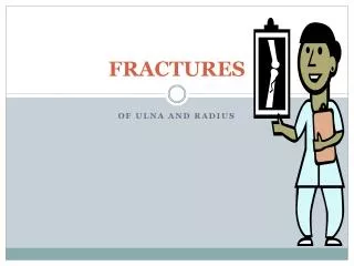
Facial and Mandibular Fractures. Presented by M.A. Kaeser, DC Spring 2009. Basic Facial Series. Three films Waters view – PA view with cephalad angulation This is the most consistently helpful view in facial trauma Caldwell view – PA view Lateral view A fourth film may be warranted

E N D
Facial and Mandibular Fractures Presented by M.A. Kaeser, DC Spring 2009
Basic Facial Series • Three films • Waters view – PA view with cephalad angulation • This is the most consistently helpful view in facial trauma • Caldwell view – PA view • Lateral view A fourth film may be warranted Submentovertex view – through the foramen magnum
Simple Rules • Look at orbits carefully • 60-70% of all facial fractures involve the orbit • Know the most common patterns of facial fractures and look for them • Bilateral symmetry can be very helpful • Carefully trace along the lines of Dolan when examining the Waters view in a facial series
Lines of Dolan • Three anatomic contours • The 2nd and 3rd lines together form the profile of an elephant
Direct Radiographic Signs of Facial Fractures • Nonanatomic linear lucencies • Cortical defect or diastatic suture • Bone fragments overlapping causing a “double-density” • Asymmetry of face
Indirect Radiographic Signs of Facial Fractures • Soft tissue swelling • Periorbital or intracranial air • Fluid in a paranasal sinus
MOIs • Auto accidents – 70% of auto accidents produce some type of facial injury (most are limited to soft tissue) • Fights/Assaults • Falls • Sports • Industrial Accidents • Gunshot Wounds *Less than 10% of all facial fractures occur in children
Fracture Types and Prevalence • Zygomaticomaxillary complex – AKA Tripod fracture = 40% • LeFort I = 15% • LeFort II = 10% • LeFort III = 10% • Zygomatic arch = 10% • Alveolar process of maxilla = 5% • Smash Fractures = 5% • Other = 5%
Tripod Fracture • Most common facial fracture • Usually occurs as a diastasis of the zygomaticofrontal suture
LeFort Fractures • Complex, bilateral fracures associated with a large unstable fragment • Involve the pterygoid plates
Three Main Planes of Weakness in the Face • Maxillary Plane • Between the maxillary floor and the orbital floor • Subzygomatic or Pyramidal Plane • MOI = down ward blow to the nasal area • Craniofacial Plane • Uncommon as an isolated injury • Occurs in association with severe skull and brain injuries
Zygomatic Arch Fracture • Usually due to a blow from the side of the face • Cause flatness of the lateral cheek area, inability to open mouth
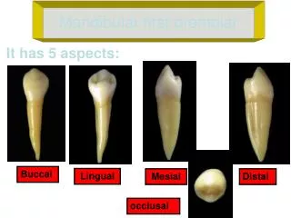
Alveolar Process of Maxilla • Associated with several fractured teeth • Chest film should be taken if all teeth are not accounted for
Smash Fracture • Severe comminution of the face • Underlying skull injury is likely
Blowout Fracture • MOI – blow to the eye, forces are transmitted by the soft tissues of the orbit downward to the thin floor of the orbit • Symptoms – enophthalmos and diplopia (usually an upward gaze) • 24% are associated with ocular injury
Nasal Bone Fracture • Most commonly missed facial fracture • Most frequently injured facial structure • Most nasal bone fractures will run perpendicular to the bridge of the nose • May be associated with more extensive injuries • Orbital rim or floor • Ethmoid or frontal sinuses
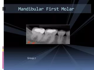
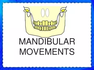
Mandibular Fractures • Clinical findings • Facial distortion • Malocclusion of the teeth • Abnormal mobility of portions of the mandible or teeth
Ring Bone Rule – AKA Pretzel-Bagel Spectrum • If you see a fracture or dislocation in a ring bone or ring bone equivalent, look for another fracture or dislocation
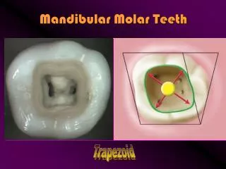
Common Sites of Mandibular Fractures and Prevalence • Body 30-40% • Angle 25-31% • Condyle 15-17% • Symphysis 7-15% • Ramus 3-9% • Alveolar 2-4% • Coronoid Process 1-2%
Double Mandibular Fractures • Usually contralateral sides of the symphysis • Common combinations include: • Angle plus the contralateral body or condyle
Mandibular Dislocation • May occur spontaneously during a large yawn • Considerable pain • Condyle (c) is anterior to the articular eminence (e)
Important Thoughts About Mandibular Fractures • Remember the ring bone rule • Symphyseal fractures can be hard to see • Panorex view provides the best single view of the mandible • Look carefully along the cortical margin of the whole mandible for discontinuities • Carefully examine the mandibular canal for discontinuities • Pathologic fractures can occur in the mandible – look for tumors or abscesses