Nervous system
790 likes | 1.82k Vues
Nervous system. by Dr. Dalia Mohamed Ali. Divisions of the Nervous System. The nervous system (N.S) is divided into two main parts : 1) The central nervous system(C.N.S ) 2) The peripheral nervous system(P.N.S). The peripheral nervous system.

Nervous system
E N D
Presentation Transcript
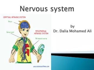
Nervous system by Dr. Dalia Mohamed Ali
Divisions of the Nervous System • The nervous system (N.S) is divided into two main parts: • 1) The central nervous system(C.N.S) • 2) The peripheral nervous system(P.N.S)
The peripheral nervous system • It is formed of: • Cranial nerves arise from brain • Spinal nerves arise from spinal cord • It is divided functionally into : • a-Somatic nervous system: • controls skeletal muscles . • b-Autonomic nervous system :Controls smooth muscle ,heart &glands (sympathetic & parasympathetic) .
The somatic nervous system is concerned with somatic functions. • It includes the nerves supplying the skeletal muscles. • Controls the voluntary movements of the body by acting on the skeletal muscles . • The autonomic nervous system is concenrned with regulation of visceral or vegetative functions. • involuntary nervous system. • The autonomic nervous system consists of two divisions: * sympathetic division * parasympathetic division. 1-somatic nervous system 2-Autonomic nervous system
Central nervous system • It includes: -Brain -Spinal cord • It is formed of: -Highly specialized nerve cells (neurons) -Supporting cells (neuralgia) • The structures of brain and spinal cord are arranged in two layers: the gray matter and white matter. • In brain the white matter is centrally placed and gray matter is in the outer part. • In spinal cord white matter is in the outer part and gray matter is in inner part.
Brain is situated in the skull. • It is continued as spinal cord in the vertebral canal through the foramen magnum of the skull bone. • Brain and spinal cord are surrounded by three layers of meninges: • the outer dura mater • middle arachnoid • inner pia mater. • The space between the arachnoid mater and pia mater is known as subarachnoid space. • This space is filled with a fluid called cerebrospinal fluid (CSF). • The brain and spinal cord are actually suspended in CSF.
A) The brain • It is formed of : • a) cerebrum ( the 2 cerebral hemispheres + the interbrain). • b) Brain stem: which includes: • Midbrain ( upper part) • Pons (middle part) • medulla oblongata (lower part) • c)cerebellum
Neuron & Neurolgia • The functional & structural unit of the Nervous System is the neuron. • Neuron is called the nerve cell • Nerve cell body: • Irregular in shape • mass of cytoplasm called neuroplasm which is covered by a cell membrane. • The cytoplasm contains a large nucleus, Nissel bodies, neurofibrils, mitochondria and Golgi apparatus. • Nissel bodies and neurofibrils are found only in nerve cell and not in other cell
It is different from other cells in: • It has branches or processes • It does not have centrosome (it cant divide) • two types of processes: 1) the axon the axons(nerve fibers) runs in groups forming the different nerves of the body. 2)Dendrites
Organization of nerve : • Many axons together form a bundle called fasciculus. • many fasciculi together form a nerve.
Myelin sheath • myelin sheath is a thick lipoprotein sheath that insulates the nerve fiber. • it is absent at regular intervals. • myelin sheath is responsible for the white color of the nerve fibers -Myelinated nerve fiber The nerve fibers which are insulated by myelin sheath. -Nonmyelinated nerve fiber The nerve fiber is not covered by myelin sheath. Function of Myelin sheath: 1- Faster conduction faster conduction of impulse 2- Insulating capacityrestricts the nerve impulse within the single nerve fiber
Classification of neuron I- depending on number of poles: 1- unipolar neurons have only one pole from which both the axon and dendrite aries 2- Bipolar neurons have two poles.axon arises from one pole and dendrites aries from the other pole 3- Multipolar neurons have many poles. One of the poles gives rise to the axon and all the other poles give rise to dendrites
II- depending on function: 1- Motor neurons (efferent) • motor neurons or efferent nueurons which carry the motor impulses from central nervous system to the peripheral effector organs like muscles,glands,blood vessels. 2- Sensory neurons (afferent) • sensory neurons or afferent neurons which carry the sensory impulses from periphery to the central nervous system.
III-depending on length of axon: 1- Golgi type I neurons (long axons) • have long axons. • The cell body of these neurons is in central nervous system and their axons reach the remote peripheral organs. 2- Golgi type II neurons (short axons) • have short axons. • These neurons are present in cerebral cortex and spinal cord.
Classification of nerve fibers: • I- depending on structure: -Myelinated nerve fibers -NonMyelinated nerve fibers • II-depending on distribution -somatic nerve fibers -autonomic nerve fibers • III-depending on origin: -cranial nerve: arise from brain -spinal nerve: arise from spinal cord
IV-depending on function: 1-sensory or afferent nerve fibers which carry sensory impulses from different parts of the body to the central nervous system 2-motor or efferent nerve fibers which carry motor impulses from central nervous system to different parts of the body. • V-depending on neurotransmitter: • Adrenergic nerve fibers that secrete noradrenaline • cholinergic nerve fibers that secrete acetylcholine
Properties of nerve fiber • Excitability • Conductivity • Refractory period: -absolute RP -relative RP • Adaptation • Summation • Infatigability
Excitability the physiochemical change that occurs in a tissue when a stimulus is applied • Conductivity the ability of nerve fibers to transmit the impulse from the area of stimulation to the other areas • Refractory period the period at which the nerve does not give any response to a stimulus -absolute RP -relative RP
Adaptation • While stimulating a nerve fiber the excitability of the nerve fiber is greater in the beginning. • continuous stimulation Later the response decreases slowly and finally the nerve fiber does not show any response at all • Infatigability • A nerve fiber cannot be fatigued, even if it is stimulated continuously for a long time • Summation • When one subliminal stimulus is applied it does not produce any response in the nerve fiber . • if two or more subliminal stimuli are applied within a short interval the response is produced
Neuroglia • Neuroglia or glia cell is supporting cells • It is nonexcitable not transmit impulses • Classification: 1- central glia cells: -astrocytes -microglia -oligodendrocytes 2- peripheral glia cells: -schwan cells -satellite cells
Receptors • It is sensory nerve ending that terminate in the periphery • Classification: • I- Exteroceptors: -cutaneous receptors: pain, pressure, touch -chemoreceptors: taste, smell -telereceptors: vision, hearing • II- Interoceptors: -viscroceptors -proprioceptors
Exteroceptors: • are the receptors which give response to stimuli arising from outside the body. 1_cutaneous receptor: • The receptor situated in the skin • also called mechanoreceptors their response to mechanical stimuli such as touch, pressure &pain. 2_chemoreceptors: • The receptors which give response to chemical stimuli are called to chemoreceptors. 3_telereceptos: • Are the receptors that give response to stimuli arising away from the body.
Interoceptors • Are the receptors which give response to stimuli arising from within the body interoceptors are of 2 types: 1-visceroceptors: • situated the viscera. 2-proprioceptors: • Are the receptors which give to change in the position of different parts of the body.
Properties of receptors • 1-specificity of response • 2-adaptation • 3-response to increase in strength of stimulus • 4-sensory transduction • 5- receptor potential
Properties of receptors 1-specificity of response • the response given by a particular types of receptor to a specific sensation. 2-adaptation • It is the decrease in the response when a receptor is stimulated continuously with constant strength.
3-response to increase in strength of stimulus • if the response given by the receptor is to be doubled the strength of stimulus must be increased 100 times. 4-sensory transduction • The process by which the energy (stimulus) is converted into electrical impulses. 5- receptor potential • Is a nonpropagatedtransmembrane potential difference that develops when a receptor is stimulated • short lived • transient • receptor potential is not action potential.
Synapse Synapse: • It is the junction between the two neurons. Classification: I- Anatomical: 1-axaoaxnic synapse • in which axon of one neuron terminates on axon of another neuron. 2-axodendritic synapse • in which axon of one neuron terminates on dendrite of another neuron. 3-axosomatic synapse • in which axon of one neuron ends on soma(cell body)of another neuron.
II- Functional: • classified into 2 types: 1-electrical synapse: • Between the presynaptic and the postsynaptic neurons. • there is direct exchange of ions between the 2 neurons though the gap junction. 2-chemical synapse: • Junction between a nerve fiber and a muscle fiber or between 2 nerve fibers • through which the signals are transmitted by the release of chemical transmitter.
Function of synapse: 1-excitatory synapses which transmit the impulses from one neuron to another. 2-inhibitory synapsesinhibit transmission of impulses. - Postsynaptic inhibition -presynaptic inhibition -renshaw cell inhibition
Excitatory synapse: • Excitatory synapse transmits the impulses from presynaptic neuron to postsynaptic neuron by the development of excitatory postsynaptic potential (EPSP). Excitatory prostsynaptic potential: • Excitatory postsynaotic potential (EPSP) is the nonptopagated electrical potential that develops during the process of synaptic transmission • the common neurotransmitter in synapse is acetylcholine.
Inhibitory synapse: Inhibition of synaptic transmission is classified into 3 types: 1-postsynaptic inhibition. 2-presynaptic inhibition. 3-renshaw cell inhibiton
Inhibitory synapse: 1- postsynaptic inhibition • due to the release of an inhibitory neurotransmitter from presynaptic terminal instead of an excitatory neurotransmitter. • the inhibitory nurotranmittrs are: gamma amino butyric acid (GABA), dopamine and glycine. • the transmitter receptor complex opens the ligand gated potassium channels instead of sodium channels. • now the potassium ions which are available in the cell body of postsynaptic neuron move to ECF . • simultaneously chloride channels also opened. • the exit of potassium ions and influx of chloride ions cause more negativity inside leading to hyperpolarization.
2- Presynaptic inhibition: • It's the synaptic inhibition which occurs because of the failure of presynaptic axon terminal to release the excitatory neurotransmitter substance (indirect inhibition). 3- renshaw cell inhibition: • It's the type of synaptic inhibition which is caused by renshaw cells in spinal cord. • the renshaw cell is stimulated it sends inhibitory impulses to a motor neurons so that the discharge from a motor neurons is reduced.
significance of synaptic inhibition • the synaptic inhibition in CNS limits the number of impulses going to muscles and enables the muscles to act properly and appropriately
Properties of synapse 1-one way conduction : • Impulses are transmitted only in one direction (from presynaptic neuron to postsynaptic neuron). 2-the synaptic delay: • Synaptic delay is a short delay that occurs during the transmission of impulses through the synapse it's due to the time taken for: • release of neurotransmitter. • passage of neurotransmitter from axon terminal to postsynaptic membrane. • action of the neurotransmitter to open the ionic channels in postsynaptic membrane.


