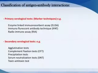
PRIMARY SEROLOGICAL TEST
PRIMARY SEROLOGICAL TEST. IMMUNOFLUORESCENT TES T. Introduction. Immunofluorescence is a serological test where the labeling of antibodies or antigens is done with fluorescent dyes ( fluorochromes).


PRIMARY SEROLOGICAL TEST
E N D
Presentation Transcript
PRIMARY SEROLOGICAL TEST IMMUNOFLUORESCENT TEST
Introduction • Immunofluorescence is a serological test where the labeling of antibodies or antigens is done with fluorescent dyes ( fluorochromes). • Fluorochromes are dyes which have the ability to absorb the short wavelength UV radiation and emit light of longer wavelength fluorescence ( visible green light). • Examples : FITC, Rhodamine , Acridine orange
Immunofluorescence - Most commonly used fluorescent dyes are: Fluorescein and Rhodamine, but other highlyfluorescent substances (Phycoerythrin and Phycobiliproteins) have also come into use. - Fluorescein: absorbs blue light and emits an intense yellow-green fluorescence - Rhodamine: absorbs yellow-green range and emits a deep red fluorescence - Phycoerythrin: efficient absorbers of light and brilliant emitters of red fluorescence
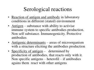
Assumptions that are important for 1 serological tests • One of reactants (Ag ,Ab) must be rendered insoluble (fixed ) on solid phase such as slide or microtiter well or glass bead. • The other reactant is conjugated to a labeling material( enzyme, fluorochrome, radioactive substance) without loss of activity.
There are two ways of doing IF staining • Direct immunofluorescence • Indirect immunofluorescence • Direct immunofluorescence Use: Direct detection of Pathogens or their Ag’s in tissues or in pathological samples • Ag is fixed on the slide • Fluorescein labeled Ab’s are layered over it • Slide is washed to remove unattached Ab’s • Examined under UV light in an fluorescent microscope • The site where the Ab attaches to its specific Ag will show apple green fluorescence
Indirect immunofluorescence: • Indirect test is a double-layer technique • The unlabelled antibody is applied directly to the tissue substrate • Treated with a fluorochrome-conjugated anti-immunoglobulin serum
Advantage over direct IF • Because several fluorescent anti-immunoglobulins can bind to each antibody present in the first layer, the fluorescence is brighter than the direct test. • It is also more time-efficient since it is only one signal labeled reagent, the anti-immunoglobulin, is prepared during the lengthy conjugation process
Indirect Elisa for detection of ANAPrinciple ( Diagram) Incubation period followed by washing FITC Anti human IgG conjugated to FITC Non specific AB Incubation period followed by washing Patient serum Ag Ag Nucleated tissues Solid phase :slide Solid phase :slide
Indirect IFA for detection of ANA in patient sample • Serum sample must be diluted 1:40 to dilute non specific Ab due to aging or autoimmune diseases such as RA. • Nucleated tissues(Ag) are immobilized on slide • Diluted patient serum is added(25ul),the slide is put in a humid chamber. • After addition of patient serum ,there is an incubation period followed by washing(3-4).
Indirect IFA for detection of ANA in patient sample • Conjugate is added. • After addition of conjugate , there is an incubation period followed by washing. • Read using Fluorescent microscope. • UV light is produced by Fluorescent microscope special lamp .This microscope must have barrier filter to protect observer’s eye from UV light.
Indirect IFA for detection of ANA in patient sample • Washing is an important step to remove unbound ( excess/ non specific)Ag or Ab • Insufficient washing results in false +ve results • Excessive washing results in false –ve results • Patient serum must be diluted 1:40 before performing test(why)
Indirect IFA for detection of ANA in patient sample(+ve/x40)
Indirect IFA for detection of ANA in patient sample (+ve/x40)
Indirect IFA for detection of ANA in patient sample (+ve/x20)
Indirect IFA for detection of ANA in patient sample (-ve/x40)
False +ve and False -ve results • False +ve • Cross reacting Ab • Presence of Rheumatoid factor or other autoimmune disease • Use of non specific Ag • Improper washing • Aging • False –ve • Improper Immune system • Blocking Ab(compete for Ag binding site of Ab) • Prozone phenomenon • Excessive washing