Download
1 / 80
800 likes | 1.03k Vues
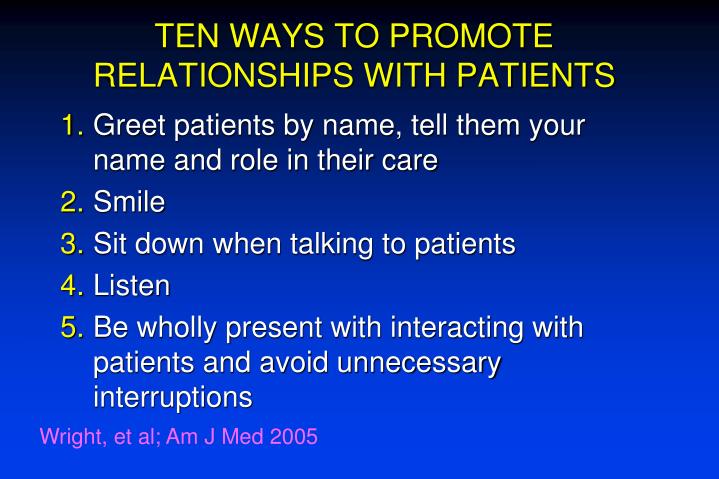
TEN WAYS TO PROMOTE RELATIONSHIPS WITH PATIENTS. Greet patients by name, tell them your name and role in their care Smile Sit down when talking to patients Listen Be wholly present with interacting with patients and avoid unnecessary interruptions. Wright, et al; Am J Med 2005.

E N D
TEN WAYS TO PROMOTE RELATIONSHIPS WITH PATIENTS • Greet patients by name, tell them your name and role in their care • Smile • Sit down when talking to patients • Listen • Be wholly present with interacting with patients and avoid unnecessary interruptions Wright, et al; Am J Med 2005
TEN WAYS TO PROMOTE RELATIONSHIPS WITH PATIENTS • Learn who your patients are and consider sharing something about yourself with them • Show the utmost respect for all patients • Be humanistic, compassionate, and caring • Even if it is a struggle to think positively of a patient, always speak of them in a positive way; this will influence your thinking positively • If you are feeling negative emotions towards a patient, try to understand why you are feeling this way Wright, et al; Am J Med 2005
The most common metabolic liver disease in children is: • A) Alpha-1 Antitrypsin deficiency • B) Hemochromatosis • C) Wilson’s Disease • D) Cystic Fibrosis • E) Familial IntrahepaticCholestasis
The most common metabolic liver disease in children is: • A) Alpha-1 Antitrypsin deficiency • B) Hemochromatosis • C) Wilson’s Disease • D) Cystic Fibrosis • E) Familial IntrahepaticCholestasis
The least common metabolic liver disease in children is: • A) Alpha-1 Antitrypsin deficiency • B) Hemochromatosis • C) Wilson’s Disease • D) Cystic Fibrosis • E) Familial IntrahepaticCholestasis
The least common metabolic liver disease in children is: • A) Alpha-1 Antitrypsin deficiency • B) Hemochromatosis • C) Wilson’s Disease • D) Cystic Fibrosis • E) Familial IntrahepaticCholestasis
Which of the following is not seen in HH? • A) Hepatomegaly • B) Hypogonadotropichypogonadism • C) Hypothyroidism • D) Heart Failure • E) Destructive arthritis • F) Erythrocytosis
Which of the following is not seen in HH? • A) Hepatomegaly • B) Hypogonadotropichypogonadism • C) Hypothyroidism • D) Heart Failure • E) Destructive arthritis • F) Erythrocytosis
HEREDITARY HEMOCHROMATOSISDefinition and Inheritance • Increased intestinal absorption of iron • Deposition in multiple parenchymal organs • Autosomal recessive • Common in Caucasians • homozygote frequency - 0.5% • heterozygote frequency - 10%
HFE GENE • Discovered in 1996 • Two mutations initially described (C282Y and H63D) • 60-100% of patients with HH are C282Y/C282Y • 5-10% of patients with clinically significant iron overload don’t have HFE mutations
HFE AND IRON OVERLOADSupporting evidence • Wild type and H63D protein bind to -2 microglobulin (-2 M) • expressed on the cell surface • facilitates iron uptake • C282Y protein does not bind to -2 M and is not expressed on the cell surface • HFE knock out mice and -2 M knockout mice develop HH • We still don’t know how the mutated HFE protein causes iron overload
There are 4 types of iron overload- can you rank them in terms of severity? • Type 1: HFE related HH (adult) • Type 2A: Hemojuvelin related juvenile HC • Type 2B: Hepcidin related juvenile HC • Type 3: Transferrin receptor 2 HC (adult) • Type 4: Ferroportin related iron overload (note: this is the only AD one)
HEPCIDIN • 25 aa peptide synthesized in hepatocytes • HAMP gene on chromosome 19q • Stimulated by inflammation and iron excess • Down-regulates ferroportin-mediated release from enterocytes, placenta, and macrophages • Levels increase 100X in anemia of of chronic disease Park et al. J Biol Chem 2001.
HEPCIDIN-2 • Hepcidin mRNA induced by dietary and parenteral iron overload • Anemia and hypoxia suppress hepcidin • Hepcidin KO mice develop iron overload • Over expression leads to iron deficiency • HFE KO mice and humans with HH have inappropriately low hepcidin levels Ahmad Blood Cells Mol Dis. 2002; Bridle Lancet 2003
The earliest sign of HC is: • A) elevated ferritin • B) elevated transferrin saturation • C) Impaired OGTT • D) 1st and 2nd MCP joint destruction
The earliest sign of HC is: • A) elevated ferritin • B) elevated transferrin saturation • C) Impaired OGTT • D) 1st and 2nd MCP joint destruction
30 yo asymptomatic female is referred for elevated serum iron studies obtained as part of her routine health evaluation. Iron = 180 g/dl (normal 35-145); TS% = 80% (normal 14-50%); ferritin = 80 g/L (normal 20-200). PMH and FMH are unremarkable. She consumes 3 glasses of wine per week and does not use iron supplements. Exam is normal. Repeat TS is 74%. What would you recommend? 1. No further evaluation 2. HFE gene test 3. Therapeutic phlebotomy 4. MRI of the liver to look for iron deposition 5. HFE gene test and phlebotomy
30 yo asymptomatic female is referred for elevated serum iron studies obtained as part of her routine health evaluation. Iron = 180 g/dl (normal 35-145); TS% = 80% (normal 14-50%); ferritin = 80 g/L (normal 20-200). PMH and FMH are unremarkable. She consumes 3 glasses of wine per week and does not use iron supplements. Exam is normal. Repeat TS is 74%. What would you recommend? 1. No further evaluation 2. HFE gene test 3. Therapeutic phlebotomy 4. MRI of the liver to look for iron deposition 5. HFE gene test and phlebotomy
Clinical Suspicion of HH No Fasting, morning TS % > 45% Stop Yes No Repeat TS % and serum ferritin elevated Recheck in 1 year Yes Secondary iron overload? Yes No HFE gene testing Treat and recheck
WHO SHOULD BE TESTED FOR HH? • Adult 1st degree relatives of a proband (HFE) • Patients with persistent liver test abnormalities or chronic liver disease (TS%) • Patients with one or more symptoms/signs suggestive of HH (TS%) • diabetes • heart failure or cardiac arrhythmias • young patients with “DJD” • Patients with porphyria cutanea tarda (HFE)
HEREDITARY HEMOCHROMATOSISClinical Features • Iron accumulation is slow (1-2 mg excess iron per day) • Clinical manifestations often do not occur until 5th decade • Clinical manifestations in females may be mild and/or delayed • Alcohol, hepatitis C, and other ill-defined environmental and genetic factors may influence disease progression
A 30 yo asymptomatic female is referred for elevated serum iron studies obtained as part of her routine health evaluation. Iron = 180 g/dl (normal 35-145); TS% = 80% (normal 14-50%); ferritin = 80 g/L (normal 20-200). PMH and FMH are unremarkable. She consumes 3 glasses of wine per week and does not use iron supplements. Exam is normal. Repeat TS is 74%. She is homozygous for C282Y. She asks about treatment. What would you recommend? 1. Phlebotomy 2. Deferoxamine 3. No treatment but follow iron tests 4. A “low iron” diet
A 30 yo asymptomatic female is referred for elevated serum iron studies obtained as part of her routine health evaluation. Iron = 180 g/dl (normal 35-145); TS% = 80% (normal 14-50%); ferritin = 80 g/L (normal 20-200). PMH and FMH are unremarkable. She consumes 3 glasses of wine per week and does not use iron supplements. Exam is normal. Repeat TS is 74%. She is homozygous for C282Y. She asks about treatment. What would you recommend? 1. Phlebotomy 2. Deferoxamine 3. No treatment but follow iron tests 4. A “low iron” diet
Author N C282Y homozygosity Elevated ferritin TS > 45% Burt 1064 1:213 60% 100% McDonnell 1653 1:276 50% 67% Adams 5211 1:327 19% 75% Olynk 3011 1:188 75% 94% Beutler 41,038 1:270 65% 57%* POPULATION SCREENING STUDIESGenotype and Phenotype *TS% > 50%
A 30 yo asymptomatic female is referred for elevated serum iron studies obtained as part of her routine health evaluation. Iron = 180 g/dl (normal 35-145); TS% = 80% (normal 14-50%); ferritin = 80 g/L (normal 20-200). PMH and FMH are unremarkable. She consumes 3 glasses of wine per week and does not use iron supplements. Exam is normal. Repeat TS is 74%. She is homozygous for C282Y. She inquires about screening her brothers and daughter. What would you suggest? 1. No screening necessary 2. HFE gene test for both 3. Transferrin saturation for both 4. HFE gene test for her brothers
A 30 yo asymptomatic female is referred for elevated serum iron studies obtained as part of her routine health evaluation. Iron = 180 g/dl (normal 35-145); TS% = 80% (normal 14-50%); ferritin = 80 g/L (normal 20-200). PMH and FMH are unremarkable. She consumes 3 glasses of wine per week and does not use iron supplements. Exam is normal. Repeat TS is 74%. She is homozygous for C282Y. She inquires about screening her brothers and daughter. What would you suggest? 1. No screening necessary 2. HFE gene test for both 3. Transferrin saturation for both 4. HFE gene test for her brothers
Family History of C282Y Homozygous HH No C282Y Mutation HFE Gene Test (adults only) C282Y homozygote C282Y Heterozygote Serum TS % and Ferritin No further evaluation Serum TS % and Ferritin Ferritin > 1000 g/L and/or elevated AST Ferritin normal Normal Ferritin elevated but < 1000 g/L and normal AST Increased Repeat ferritin annually Follow and consider liver biopsy or trial of quantitative phlebotomy if ferritin is >500 g/L Phlebotomy Liver biopsy and phlebotomy
One of her brothers is homozygous for C282Y. His serum iron is 220 g/dl, TS=88% and ferritin 575 g/L. His liver tests are normal. He is healthy and asymptomatic. He consumes 1 alcoholic beverage per day. What would you recommend? • 1. Liver biopsy followed by phlebotomy • 2. Phlebotomy • 3. MRI to assess hepatic iron stores • 4. Stop alcohol and repeat iron tests in 3 mos • 5. Observation
One of her brothers is homozygous for C282Y. His serum iron is 220 g/dl, TS=88% and ferritin 575 g/L. His liver tests are normal. He is healthy and asymptomatic. He consumes 1 alcoholic beverage per day. What would you recommend? • 1. Liver biopsy followed by phlebotomy • 2. Phlebotomy • 3. MRI to assess hepatic iron stores • 4. Stop alcohol and repeat iron tests in 3 mos • 5. Observation
LIVER BIOPSY IN HEMOCHROMATOSIS • Rationale • confirm diagnosis • exclude cirrhosis • Liver biopsy not needed to confirm diagnosis of HH in patients homozygous for C282Y with an elevated transferrin saturation and ferritin. • Cirrhosis rare in C282Y homozygotes with ferritin < 1000 g/L and normal aminotransferases.
In a patient with HH and cirrhosis, the risk of HCC is • A) Higher than that of viral hepatitis • B) Lower than viral but higher than NASH • C) Lower than NASH, but higher than cholestatic diseases • D) very low
In a patient with HH and cirrhosis, the risk of HCC is • A) Higher than that of viral hepatitis • B) Lower than viral but higher than NASH • C) Lower than NASH, but higher than cholestatic diseases • D) very low
HEMOCHROMATOSIS AND CIRRHOSIS • Major predictor of survival • Complications of ESLD uncommon • Death usually due to liver cancer • 30% with HH cirrhosis develop HCC • 200X increased risk • Risk persists despite iron depletion • HCC rare in the absence of cirrhosis
Clinical suspicion of HH No Fasting, morning TS% >45% Stop Yes No Repeat TS% and ferritin > normal Recheck in 1 year Yes Yes Secondary iron overload? Treat underlying cause No HFE gene testing; C282Y homozygote? No Yes 1. Ferritin <1,000 ug/L 2. Normal AST Yes No Phlebotomy Liver biopsy histology and HIC consistent with HH? Yes No Phlebotomy Follow CP1134735-1
HEREDITARY HEMOCHROMATOSISTreatment • Treat HH patients with an elevated ferritin • Phlebotomy - the preferred treatment modality • 500 ml per week • 500 ml blood = 250 mg iron • Goal ferritin < 50 ug/L +/- TS% < 50% • Maintenance approximately every 3 months • Desferoxamine- infrequently used
18 16 14 12 10 20 8 6 4 2 0 Response of Transferrin Saturation and Serum Ferritin to Phlebotomy 25 2,500 100 20 2,000 80 15 1,500 60 Cumulative iron removal (g) Transferrin saturation (%) Serum ferritin (ng/mL) 10 1,000 40 5 500 20 0 0 0 Months Hepatology A Textbook of Liver Disease, ed. Zakim and Boyer CP1134735-7
Pituitary GonadotropinsMSH • Hypothyroidism • Cardiomyopathy • Conduction disorders • Diabetes mellitus • Testicular atrophy • Impotence HEMOCHROMATOSIS • Increased skin pigmentation • Increased liverbiochemistries • Cirrhosis • Hepatocellular carcinoma • Arthropathy(m-p joints)Chondrocalcinosis Blue=reversible, yellow=usually not reversible; Modified from AGA clinical teaching project CP1134735-8
HEREDITARY HEMOCHROMATOSISOther management • Avoid • ETOH • iron and vitamin C supplements • raw shellfish • A “low iron diet” is not necessary • Vaccinate against hepatitis A and B
A 35 yo female is found to have an elevated serum ferritin. Serum iron 120 g/dl, TS%=35%, ferritin=814 g/L. PMH: Hepatitis C Meds: MVI with iron (for 1 year) PE: Normal Labs: Hgb= 11 g/dl. AST=95, ALT= 134, the rest of her liver tests are normal. What is the most likely explanation for her abnormal serum iron studies? 1. Hereditary hemochromatosis 2. Anemia 3. Hepatitis C 4. Excess iron supplementation
A 35 yo female is found to have an elevated serum ferritin. Serum iron 120 g/dl, TS%=35%, ferritin=814 g/L. PMH: Hepatitis C Meds: MVI with iron (for 1 year) PE: Normal Labs: Hgb= 11 g/dl. AST=95, ALT= 134, the rest of her liver tests are normal. What is the most likely explanation for her abnormal serum iron studies? 1. Hereditary hemochromatosis 2. Anemia 3. Hepatitis C 4. Excess iron supplementation
Differential Diagnosis of an Isolated Elevated Serum Ferritin • Infection, inflammation or malignancy • Chronic liver disease • Ferroportin disease • Aceruloplasminemia (rare) • Hereditary hemochromatosis (rare) • Hyperferritinemia cataract syndrome
FERROPORTIN DISEASE • First reported in 1999 • SLC40A1 gene on chromosome 2q • Iron exporter (enterocyte, placenta, liver and macrophage) • Autosomal dominant iron overload disorder • The second most common inherited form of iron overload