Download
1 / 111
1.23k likes | 2.45k Vues
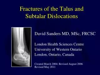
Fractures of the Talus and Subtalar Dislocations. David Sanders MD, MSc, FRCSC London Health Sciences Centre University of Western Ontario London, Ontario, Canada Created March 2004; Revised August 2006 Revised May 2011. Outline:. Talar Neck Fractures Anatomy Incidence Imaging

E N D
Fractures of the Talus and Subtalar Dislocations David Sanders MD, MSc, FRCSC London Health Sciences Centre University of Western Ontario London, Ontario, Canada Created March 2004; Revised August 2006Revised May 2011
Outline: Talar Neck Fractures Anatomy Incidence Imaging Classification Management Complications Talar body, head and process fractures Subtalar dislocations Classification Management Outcomes
Anatomy • Surface 60% cartilage • No muscular insertions
Blood Supply 4 primary arterial sources: • Artery of tarsal canal • Artery of tarsal sinus • Dorsal neck vessels • Deltoid branches lateral medial Inferior view of talus, showing vascular anastomosis
Vascularity • Artery of tarsal canal supplies majority of talar body Top View Side View DeltoidBranches Superior Neck Vessels Artery of Tarsal Canal Posterior tubercle vessels Artery of Tarsal Canal Superior Neck Vessels Posterior tubercle vessels Artery of Tarsal Sinus Artery of Tarsal Sinus
Vascularity Talus ORIF Technique
Incidence • 2 % of all fractures • 6-8% of foot fractures • High complication rates • avascular necrosis • post-traumatic arthritis • malunion
Mechanism of Injury • Hyperdorsiflexion of the foot on the leg • Neck of talus impinges against anterior distal tibia, causing neck fracture • If force continues: • talar body dislocates posteromedial • often around deltoid ligament
Injury Mechanism • Previously called “aviator’s astragalus” • Usually due to motor vehicle accident or falls from height • Approximately 50 % of patients have multiple traumatic injuries
Biomechanics • Theoretical shear force across talar neck: • 1200 N during active motion [Swanson 1992] Fracture fixation must withstand this force to permit active motion in the postoperative phase
Imaging Complex 3-D structure Multiple plain film orientations: AP, Lateral, Broden, & mortise views demonstrates joint congruity of ankle and subtalar joint Canale view for longitudinal alignment: approximately 15° IR to get calcaneous out of view Canale View
Canale View • Slight ankle plantarflexion with knee bent to rest foot on the table • 15 degree pronation • Xray Tube • 15 degree from vertical Canale View
CT Scan • Most useful assessment tool for surgical planning • Confirms displacement • Demonstrates subtalar joint reduction, comminution, osteochondral fractures/debris
MRI Scan • Primary role in talus injuries is to assess complications, especially avascular necrosis • May be poor quality if extensive hardware present Zone of osteonecrosis following distribution of Artery of Tarsal Canal
Talar Neck Fractures: Classification • Hawkins, 1970 • Predictive of AVN rate • Widely used
Hawkins ClassificationHawkins, LR, JBJS, 52A: 991, 1970 • Nondisplaced <10% • Subtalar Displacement <40% • Subtalar & TC ~90% • *Pantalar 100% Talus ORIF Technique *Canale, ST, JBJS, 60A: 143, 1978
Hawkins 1 Type I: undisplaced AVN rate 0 – 13 % Uncommon as most talar neck fractures are displaced
Hawkins 2 Displaced fracture with subtalar subluxation / dislocation A) fracture line enters subtalar joint B) subtalar joint intact AVN 20 – 50 % Most common type
Hawkins 3 Subtalar and ankle joint dislocated Talar body extrudes, usually around deltoid ligament Commonly open fractures, reduction very difficult Closed: plantar flex foot & flex knee Open: joy sticks AVN 83 – 100 %
Hawkins 4 Added to classification by Canale, 1974 Incorporates talonavicular subluxation Rare variant Complex talar neck fractures which otherwise do not fit classification are included as Type 4 injuries
Classification: • Comminution: • An important additional predictor of results, especially regarding prognosis re: • Malunion • Subtalar joint arthritis • Included in AO-OTA classification as a modifier
Goals of Management • Immediate reduction of dislocated joints • Vascularity • Cutaneous tension • Vascular compromise • Anatomic fracture reduction • Stable fixation • Facilitate union • Avoid complications
Definitive Treatment • Prompt anatomic operative reduction • “die is cast” with injury • Use “joy stick” K-wires for reduction • Articular bone affects ROM (V, Sup, PF) • Maintenance of reduction with HW • Screws (AP, PA), countersink • Plates (2.0 mini condylar or T) • Dual incisions • Do not dissect capsular attachments • Maintain as much soft tissue attachments Talus ORIF Technique
Treatment of Talar Neck Fractures • Emergent reduction of dislocated joints • Stable internal fixation • Debridement of subtalar joint • Maintain as much soft tissue/vascular attachments • Choice of fixation and approach depends upon personality of fracture
Treatment of Talar Neck Fractures • Post operative rehabilitation: • Sample protocol: • Initial immobilization, 2-6 weeks depending upon soft tissue injury and patient factors, to prevent contractures and facilitate healing • Non weight-bearing, Range of Motion therapy until 3 months or fracture union
Hawkins I Fracture Options: Non Operative & Non-Weight-Bearing Cast for 4-6 weeks followed by removable brace and motion OR: Percutaneous screw fixation and early motion
Hawkins II, III, and IV Fractures: • Results dependent upon development of complications • Osteonecrosis • Malunion • Arthritis
Case Example 29 yo male ATV rollover Isolated injury LLE
These injury films are tough to interpret, but close review demonstrates the dislocated talar body with some comminution.
Diagnosis • “Hawkins’ 3” talar neck fracture • Associated comminution, probably involving medial column and subtalar joint
Controversies for this Case: ???? • Surgical timing • Closed reduction • Surgical approach • Fixation
Surgical Timing • In general: emergent reduction of dislocated joints • Allow life threatening injuries to take priority and resuscitate adequately first
Closed Reduction? • May be very useful, particularly if other life threatening injuries preclude definitive surgery • Difficult in Hawkins’ 3 and 4 injuries
Closed Reduction Technique: • Adequate sedation • Flex knee to relax gastrocs • Traction on plantar flexed forefoot to realign head with body • Varus/valgus correction as necessary • Direct pressure on talar body • Adjunct: traction pin, general anesthetic, etc • Avoid repetitive reduction attempts in order to avoid skin compromise or tearing
External Fixation • Limited roles: • Multiply injured patient with talar neck fracture in whom definitive surgery will be delayed • Temporizing measure to stabilize reduced joints • Construct bridges tibia – calcaneus – midfoot
Surgical Approaches: Options • 1 incision techniques: • Anteromedial or • Anterolateral • Problem: difficult to visualize the entire talar neck and subtalar joint without significant soft tissue stripping and devascularization • Problem: usual medial comminution inhibits accurate read of fracture reduction
Surgical Approaches: Options • 2 incision technique: (generally preferred) • Anteromedial and lateral/anterolateral • Problem: 2 skin incisions, close together • Benefit: excellent fracture visualization at critical sites of reduction and subtalar joint with less stripping
1st Approach: Anteromedial Medial to TibAnt Make incision more posterior for talar body fractures to facilitate medial malleolar osteotomy
1st Approach: Anteromedial • Provides view of neck alignment and medial comminution • Extend incision distally to talonavicular joint – hardware is placed distal to proximal and needs to be well countersunk to avoid impingement
2nd Approach: Lateral • Tip of Fibula directly anterior • Mobilize EDB as sleeve • Protect sinus tarsi contents
2nd Approach: Lateral • Visualizes Anterolateral alignment and subtalar joint • Facilitates Placement of “Shoulder Screw” or lateral plate
2 incisions: Skin bridge • Narrow skin bridge but generally well tolerated • Talus fractures generally have less soft tissue problems compared to plafond or calcaneus fractures
Fixation Options • Stable Fixation to allow early motion is the goal • 1200 N stress across talar neck during early motion (Swanson JBJS 1992) • That is a lot of force! 2 A to P screws only resists about 1 kN of stress so need more fixation
Surgical Tactics: Fixation • Anterior • Partial threaded screws • Fully threaded screws • Mini-fragment plates (2.0, 2.4 mm) • Posterior • Lag screws Implant selection depends upon injury, degree of comminution, bone quality
Posterior to Anterior Fixation: 90° • Screw fixation stronger from posterior than from anteromedial (T Bray) • Screws perpendicular to fracture site • 2 PA screws in compression are able to withstand the theoretical shear force of active motion • Avoid excessive posterior capsular stripping • Creates potentially 3 incisions • (posterior, medial, lateral)
Anterior Screw Fixation: Screw fixation alone is acceptable for non-comminuted fractures, but consider adding a plate if there is comminution. • Easy to insert under direct visualization • This example: displaced type 2: 4 A-P screws including medial “buttress” fully threaded cortical screws and lateral “shoulder” screws
Anterior Screw Fixation: • This example: • 4.0 mm partially threaded compression screws through non-comminuted columns • Mini-fragment (2.4 mm) screws for osteochondral fragments • Consider Titanium for MRI
Plate Fixation: • Very useful in comminuted fractures: • 2.0 or 2.4 mm plates • Easiest to apply to lateral cortex – impinge on medial side
Hawkins 3 Talus ORIF Technique