Download
1 / 30
370 likes | 1.68k Vues
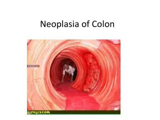
Polyps – Where do they come from and what do you do with them?!. Ron G. Landmann, MD Grand Rounds Department of Surgery St. Luke’s-Roosevelt Hospital Center March 21, 2007. Polyps. Cancer epidemiology Definition of the malignant polyp Natural history of adenomatous polyps

E N D
Polyps – Where do they come from and what do you do with them?! Ron G. Landmann, MD Grand Rounds Department of Surgery St. Luke’s-Roosevelt Hospital Center March 21, 2007
Polyps • Cancer epidemiology • Definition of the malignant polyp • Natural history of adenomatous polyps • Biology of polyps • The anatomy of the polyp • Correlations with Malignancy • Endoscopic polypectomy alone??? • Special considerations * No discussion of technique
Colorectal Cancer – Epidemiology • Incidence: Approx. 150,000 cases/year • Deaths: Approx. 50,000 deaths/year • At diagnosis • 10% in situ disease • 30% local disease • 30% regional disease • 30% distant disease • 5 year survival, all patients: 50% • local - 90% • regional - 60% • distant - 5% U.S. Cancer Statistics Working Group. United States Cancer Statistics: 2003 Incidence and Mortality (preliminary data). Atlanta (GA): Department of Health and Human Services, Centers for Disease Control and Prevention, and National Cancer Institute; 2006.
Incidence/Prevalence of Polyps • Adenomatous polyps • 30% of Western population • Most cancers arise from polyps • *excludes syndromes
Carcinoma in situ vs. cancer • Think • Carcinoma in situ = high grade dysplasia • Carcinoma in situ≠ cancer Histology Colorectal cancer is defined by invasion of/through muscularis mucosa
Histology • Colorectal cancer is defined by invasion of muscularis mucosa • Lymphatics are located in submucosa Genetic model of colorectal tumorigenesis
Relationship Between TNM Stage and Survival in Colorectal Carcinoma CA Cancer J Clin 2004;54;295-308
Treatment of CRC • Polypectomy • Colonic Resection • Treatment depends on the risk of lymph node metastasis. • Pathology is key! • Colorectal cancer is defined by invasion of muscularis mucosa • Lymphatics are located in submucosa
Incidence of malignant polyps • Definition • Malignant polyps or T1 lesions (limited to the submucosa) • Represent 5% of all adenomas • Colonoscopy polypectomy series: 2 – 12% • Colorectal resection series: 4 – 9%
Haggitt Level (1985)Classification of polyps with invasive cancer Haggitt RC, Glotzbach RE, Soffer EE, Wruble LD. Prognostic factors in colorectal carcinoma arising in adenomas: Implications for lesions removed by endoscopic polypectomy. Gastroenterology 89:328-36, 1985, p 330. Villuous/sessile (flat) polyps with invasive cancer are by definition Haggitt 4.
Sessile Polyps Kudo, 1993 • Risk of lymph node metastasis in each sessile lesion is not the same • Haggitt’s: no detail for sessile lesions • Classification of submucosal invasion: • Sm1—Invasion into the upper third of the submucosa • Sm2—Invasion into the middle third of the submucosa • Sm3—Invasion into the lower third of the submucosa • High rate of LN metastasis: 12-25%
Sm system • Able to determine Sm1, Sm2, Sm3 in 97% of cases • Haggitt Level 1, 2, 3 = Sm1 • Haggitt Level 4 = Sm1, Sm2, or Sm3 • Endoscopist must properly resect and prepare specimen • Pathologist must properly section and examine all layers
Correlations with MalignancySize Muto, 1975
Correlations with MalignancySize Muto, 1975 Nusco, 1997
Relationship betweenSize and Morphology St. Mark’s Hospital Data
Increased risk of LN Metastasis • Unfavorable pathologic features of malignant CR polyps • Poor differentiation (only on univariate) • Lymphovascular invasion (P < 0.009) • Invasion below submucosa (Haggitt Level 4) • Depth of invasion in Sm3 (P < 0.001) • Site in lower 1/3 of the rectum (P < 0.001) • Positive resection margin (< 1 mm or 1 HPF) • Not really – this is inadequate treatment, not an adverse risk factor! P-values from Nascimbeni et al. N = 353 T1 colorectal sessile lesions
Management of Pedunculated Malignant Polyps • Haggitt Level 1, 2, 3 • Complete excision or snaring • Risk of LN metastasis < 1% • Haggitt Level 4 • Treat as sessile lesions
Management of Sessile Malignant Polyps • < 2cm in diameter • Adequate snare in one piece via colonoscopy • Requires microscopic free margin of at least 2mm • Piecemeal removal • Requires further excision/follow-up or resection • High risk factors (LVI, Sm3, distal 1/3 rectum) • Oncologic resection • Full thickness transanal excision
Lesions amenable to colonoscopic polypectomy • Pedunculated or sessile < 2cm • Well/moderately differentiated • No lymphovascular invasion • Haggitt Level 1-3 or Sm1 • Close follow-up available • Endoscopically complete excision • Negative resection margins (2mm)
Criteria for Treatment of Malignant CR Polyps by Polypectomy Alone • Determined by risk of metastasis • Low risk of Lymph Node Metastasis • Pedunculated • Haggitt Level 1, 2, 3 • Level 4 Sm1, Sm2 • Sessile • Sm1, Sm2 • High risk of Lymph Node Metastasis • Lower 1/3 of the submucosa (Sm3) • LVI • Distal 1/3 of rectum
Malignant Colorectal Polyps that Should have an Oncologic Bowel Resection • Lesions in colon • Pedunculated Haggitt Level 4 with invasion into distal third of submucosa (Sm3) or LVI • Sessile lesions removed with margin < 2mm • Sessile lesions removed piecemeal • Sessile lesions with depth of invasion into distal third of submucosa (Sm3) • Sessile lesions with LVI • Lesions in middle third and upper third rectum • Same as lesions in colon • Lesions in distal third rectum • Pedunculated Haggitt Level 4 with invasion into distal third of submucosa (Sm3) or pedunculated lesions with LVI • All sessile lesions
What if ??? • What if it’s clipped in ½? • Pedunculated • Repeat endoscopy. • Require good resection with margin (2mm) • Sessile • Requires operative oncologic resection (even if Sm1, Sm2) • Unable to determine exact pathologic depth • What if it’s shredded by forceps? • Requires operative oncologic resection • What if it’s a very small lesion? • Requires marking/tattoo CIRCUMFERENTIALLY • What if it’s carcinoma in situ? • It’s not cancer. This is high grade dysplasia. Requires close follow-up. • Unless, • poor margins: repeat endoscopy with good margins • Piecemeal resection: discussion with pathologist and patient • What if it’s a large, non-endoscopically resectable polyp? • Repeat endoscopy (2nd MD?) • Oncologic resection
Other considerations… • When in doubt • Repeat colonoscopy (endoscopy) • Personally review pathology • Get a second opinion • Have a frank discussion with patient
Polyps • Natural history of adenomatous polyps • Biology of polyps • Cancer epidemiology • The anatomy of the polyp • Correlations with Malignancy • Endoscopic polypectomy alone??? • Special considerations • Indications for Polypectomy • What if it’s clipped in ½ • What if it’s shredded by forceps? • Pathology… • Marking/tattoo • Chances of Malignancy by histopath and size/morphology • * NO technique **