Download
1 / 43
790 likes | 3.69k Vues
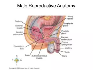
Anatomy of the Male Reproductive System. Anatomy of the Male Reproductive System. External genitalia (can be seen on the body surface) penis scrotum Internal genitalia (can’t be seen on the body surface) sperm producing organs testes ducts that move sperm from the testes out of the body

E N D
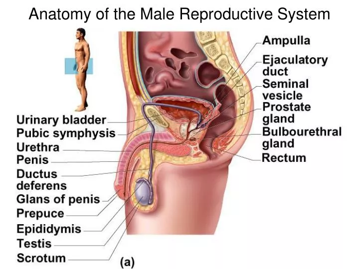
Anatomy of the Male Reproductive System • External genitalia (can be seen on the body surface) • penis • scrotum • Internal genitalia (can’t be seen on the body surface) • sperm producing organs • testes • ducts that move sperm from the testes out of the body • epididymis • vas (ductus) deferens • ejaculatory duct • urethra • exocrine glands that secrete fluids into the ducts adding to the sperm to make semen • seminal vesicles • prostate gland • bulbourethral (Cowper’s) gland
Penis • External penis consists of: • a shaft • a glans penis a prepuce (foreskin) covers the glans • Internal penis consists of: • the urethra • transports urine and semen out of the body • three cylindrical bodies of erectile tissue, a spongy network of vascular sinuses (spaces) which fill with blood during sexual excitement • Corpora cavernosa • paired erectile bodies dorsal to the urethra which are responsible for erection • Corpus spongiosum • surrounds the urethra and prevents the collapse of the urethra during erection
Male Sexual Response: Erection • Enlargement and stiffening of the penis due to the filling of erectile tissue with blood • During sexual arousal, a Parasympathetic NS reflex promotes the vasodilation of penile arteries • blood fills the erectile tissue • Expansion of the corpora cavernosa compresses the veins that drain blood out of the penis, preventing the flow of blood out of the erectile tissue
Scrotum • Sac of skin and muscle that hangs outside the abdominal cavity which hold the testes • Its external positioning keeps the testes at 34C which is required for sperm production • Two sets of muscles maintain testicular temperature: • dartos • a layer of smooth muscle deep to the skin • cremaster • smooth muscle surrounding the testes • Contraction/relaxation of both muscles raise/lower the testes toward/away from the abdominal cavity increasing/decreasing testicular temperature
Testes • Organs responsible for the production of: • male sex steroid hormone testosterone • sperm • Organized into hundreds of sperm producing seminiferous tubules • the tubules are made of a simple columnar epithelium of sertoli (Nurse) cells • the sperm development (spermatogenesis) occurs between sertoli cells from the basal surface (inside the body) of the seminiferous tubule to the lumen (outside the body) • Interstitial (Leydig) cells found between adjacent seminiferous tubules synthesize testosterone • Fluid within the seminiferous tubules flows toward the epididymis carrying the sperm
Hormonal Control of Testicular Function • The secretion of testosterone is controlled by gonadotropic releasing hormone (GnRH) and leutenizing hormone (LH) secreted from the hypothalamus and anterior pituitary gland, respectively • LH binds to receptors on Lyedig cells which stimulates the synthesis of testosterone • Some of the testosterone leaves the testes causing effects in various locations in the body causing increase bone and muscle density; facial, axillary and genital hair growth, lengthening of vocal cords • Some of the testosterone remains in the testes to support spermatogenesis
Epididymis • A mass of coiled tubes on the superficial surface of each testis that the sperm must pass through prior to ejaculation • Sperm become mature (capable of fertilizing an ovum) as pass through its tubes toward the vas deferens • During ejaculation, a layer of smooth muscle that surrounds the distal epididymis contracts, expelling sperm into the vas deferens
Ductus (Vas) Deferens and Ejaculatory Duct • A duct that propels ejaculated sperm from the epididymis towards the urethra • Its distal end merges with the duct of the exocrine seminal vesicle to give rise to the ejaculatory duct • A vasectomy is an effective form of birth control whereby the vas deferens is cut and tied
Male Sexual Response: Ejaculation • The propulsion of semen from the male duct system is coordinated by a Sympathetic NS reflex causing: • the smooth muscle surrounding the reproductive ducts and accessory glands to contract • Semen is a fluid consisting of sperm and the exocrine secretions of the 3 accessory glands: • Seminal vesicles • Prostate gland • Bulbourethral glands
Seminal Vesicles • 2 glands on the posterior wall of the bladder • Secrete seminal fluid into ejaculatory duct • accounts for 60% of semen volume • alkaline • neutralizes the acidic environment of the vagina • contains fructose • energy source for the sperm • Sperm and seminal fluid mix in the ejaculatory duct and move towards the urethra
Prostate Gland • Doughnut-shaped gland that encircles the urethra just inferior to the bladder secretes prostate fluid into the urethra • accounts for 33% of semen volume • contains enzymes that stimulate sperm movement in the vagina
Bulbourethral Glands (Cowper’s Glands) • 2 pea-sized glands inferior to the prostate • Produce thick, clear mucus during erection which: • neutralizes traces of acidic urine in the urethra • lubricates the urethra to facilitate the ejaculation of semen
Anatomy of the Female Reproductive System • External genitalia (collectively referred to as vulva) • labia majora • labia minora • clitoris • Internal genitalia • ova producing organs • ovaries • ducts/organs • uterine (fallopian) tubes • uterus • vagina
Ovaries • Located on either side of the uterus • Located in superficial surface of the ovary (cortex) are primordialfollicles (200,000 per ovary) • Each primordial follicle consists of: • an oocyte (immature egg cell) surrounded by a single layer of follicular cells • Every 28 days the pituitary hormones FSH and LH stimulate the growth of one follicle and maturation of the oocyte within the follicle called the ovarian cycle
Uterine (Fallopian) Tubes • Tubes lined with ciliated columnar epithelium create a flow of peritoneal fluid into the uterine tubes to “pull” the ovulated ovum into the uterine tube and moves it toward the uterus • The fertilization of an ovum by a sperm occurs within the distal ¼ of the tube (closest to the ovary)
Uterus • Hollow, thick-walled organ located in the pelvis, superior to the vagina which provides an ideal location for the implantation and 9 month development of a fertilized ovum There are 3 anatomical regions • Cervix • narrow neck which connects to the vagina inferiorly • Body • large middle portion of the uterus • Fundus • rounded superior region that connects to the 2 uterine (fallopian) tubes
Uterine Wall Composed of three layers • Endometrium • a 2 layered lining consisting of: • a superficial simple columnar epithelium • a deep loose connective tissue containing blood vessels and uterine glands(exocrine) • changes in thickness during the 28 day uterine (menstrual) cycle • Myometrium • thick middle layer consisting of smooth muscle • contracts during labor and childbirth • Perimetrium • outermost layer consisting of elastic but tough connective tissue • similar to the visceral peritoneum
Vagina Thin-walled tube lying posterior to the bladder and anterior to the rectum the organ of intercourse connects the external environment to the uterus provides a passageway for: sperm menstrual flow birth
Ovarian and Uterine (Menstrual) Cycles • Every 28 days in a non-pregnant female between puberty (first menstruation) and menopause (cessation of menstruation) there are changes that occur SIMULTANEOUSLY in one of the ovaries and the uterusof a non-pregnant female • Ovarian Cycle • controlled by GnRH, FSH and LH which target the ovaries and stimulate the secretion of estrogen and progesterone • Uterine Cycle • Estrogen and progesterone from the ovary target the endometrium (luminal wall) of the uterus causing it to grow and secrete uterine milk into the uterine lumen (exocrine)
Ovarian Cycle Follicular phase (days 1 – 14) • GnRH from the hypothalamus stimulates FSH and LH secretion from the anterior pituitary gland • FSH stimulates the mitosis (growth) of the follicular cells of a primordial follicle developing into a primary follicle • LH stimulates estrogen secretion from follicular cells • follicular cells secrete a layer of proteins around the ova called the zona pellucida • continues to grow into a secondary follicle • develops a fluid filledantrum in the center • estrogenlevels begin to rise steadily as the follicular cells continue secretion • continue growth into a Graafian (mature) follicle
Ovarian Cycle • Ovulation (day 14) • Therisinglevels estrogen secreted from the Graafian follicle causes a surge (release) of LH (and FSH) • The LH surge stimulates a rapid production of antral fluid which fills the follicle beyond capacity causing it to rupture • the ruptured follicle ejects (ovulates) the ovum (with a few layers of surrounding follicular cells called the corona radiata) into the fallopian tube • most of the follicular cells of the ruptured Graafian follicle remain in the ovary and organize into a corpus luteum
Ovarian Cycle • Luteal phase (days 14 – 28) • period of corpus luteum activity • LH from the surge: • transforms a ruptured follicle into a corpus luteum • stimulates the secretion of estrogen and progesterone from the corpus luteum • LH level decreases with time from LH surge
The Fate of Corpus Luteum Depends on LH • If fertilization of the ovulated ovum by a sperm (pregnancy) DOES NOT OCCUR, the corpus luteum degenerates into a corpus albicans (small scar) • causes a reduction in estrogen and progesterone • causes the secretion of GnRH, FSH and LH allowing the cycle to begin again • If fertilization of the ovulated ovum by a sperm (pregnancy) DOES OCCUR, the fertilized egg secretes a hormone called human chorionic gonadotropin (hCG) which stimulates the anterior pituitary gland for continued secretion of LH • prevents the degeneration of the corpus luteum
Uterine (Menstrual) Cycle • Menstrual phase (days 1 – 5) • menstrual bleeding causes the endometrium to shed (decrease thickness) • flows out of the vagina • caused by the decrease in estrogen levels from the degeneration of the corpus luteum during the previous cycle • Proliferative phase (days 6 – 14) • an increase in estrogen levels during the follicular phase stimulates endometrium growth (thickness) • increase in vascular supply and develops exocrine secreting uterine glands • Secretory phase (days 14 – 28) • uterine glands secrete uterine milk into the uterus • stimulated by an increase in progesterone levels during the luteal phase
Endometrium decreases in thickness (sheds) at the beginning of the cycle increases in thickness during the middle of the cycle secretes uterine milk at the end of the cycle that nourish a developing fertilized egg contains many blood vessels to supply a developing embryo and fetus develops into the placenta
Fertilization The first sperm to reach the ovum exocytoses the proteases of the acrosome which digest a layer of proteins around the ovum the zona pellucida surrounding the egg to gain access to the cell membrane the sperm and ovum cell membranes fuse together to create a single cell (zygote) the zygote begins a period of rapid mitotic divisions (cleavage) which increases the number of cells as it continues to travel towards the uterus
Placenta • A temporary organ made of embryonic and maternal tissue 2 layers that allows for the exchange of substances between maternal and embryonic/fetal blood without physical contact between these separate circulatory systems • At the end of the 1st trimester it takes over the secretion of estrogen and progesterone from the corpus luteum causing a rapid increase in both hormones for the remainder of pregnancy • An increase in estrogen levels in the mother stimulates: • breasts enlargement • the synthesis of oxytocin receptors on the myometrium of the uterus in preparation for labor and child birth
Lactation • During the later stages of pregnancy, estrogen and progesterone levels are very high which stimulates the hypothalamus to secrete prolactin-releasing hormone (PRH) which targets the anterior pituitary • the anterior pituitary responds by secreting prolactin • stimulates the production of milk in the alveoli in the breasts
Lactation • After birth, prolactin levels decrease due to the decline estrogen and progesterone levels • The stimulation for prolactin secretion comes from suckling (activates mechanoreceptors in breast) • Suckling has 2 main effects: • stimulate prolactin secretion to continue milk production • stimulate oxytocin secretion to stimulate contraction of smooth muscle around the alveolar ducts in the breast to move milk towards the nipple • reduces the size of the uterus by stimulating contraction