Understanding Animal Behavior and Natural Selection
Explore the terms stimulus, response, and reflex in animal behavior. Learn about the nervous system components involved in responses to stimuli and reflex arcs. Understand how natural selection influences animal responses through real-life examples. Discuss the Blackcap bird species' migration patterns as a case study for genetic control and responses to environmental changes.


Understanding Animal Behavior and Natural Selection
E N D
Presentation Transcript
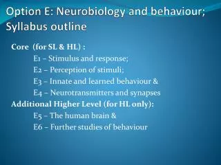
Syllabus Statements • E.1.1 Define the terms stimulus, response and reflex in the context of animal behavior. • E.1.2 Explain the role of receptors, sensory neurons, relay neurons, motor neurons, synapses and effectors in the response of animals to stimuli. • E.1.3 Draw and label a diagram of a reflex arc for a pain withdrawal reflex, including the spinal cord and its spinal nerves, the receptor cell, sensory neuron, relay neuron, motor neuron and effector. • E.1.4 Explain how animal responses can be affected by natural selection, using two examples.
Terms • Stimulus = • a change in the environment (internal or external) that is detected by a receptor and elicits a response • Response = • a change in the organism based on the stimulus that is received • Reflex = • a rapid unconscious response
Nervous systems are used in response to stimuli. Behavior is the pattern of responses in an animal.
The 5 parts of the reflex arc and their roles • 1. receptors: to detect a stimulus; receptors can be sensory cells or nerve endings of sensory neurons. • 2. sensory neurons: to receive messages across synapses, from receptors and carry them to the central nervous system (spinal cord or brain). • 3. relay neurons: to receive messages, across synapses, from sensory neurons, and pass them to the motor neurons that acan cause an appropriate response.
Reflex arc continued: • 4. motor neurons: to receive messages, across synapses, from relay neurons and carry them to an effector. • 5. effectors: to carry out a response after receiving a message from a motor neuron; effectors can be muscles, which respond by contracting, or glands, which respond by secreting.
Sequence of events in response to pain. • Nerve endings in the skin of the rabbit’s nose detect the pain caused by the stings. These cells are called pain receptors. The pain receptors are nerve endings of sensory neurons. • These sensory neurons carry impulses from the nose of the rabbit to its central nervous system. • The impulses travel to the ends of the sensory neurons where there are synapses with relay neurons. Messages are passed to the relay neurons by synaptic transmission. • The relay neurons have synapses with motor neurons, which carry impulses out of the central nervous system, to muscles in the rabbit’s body. • 5. Messages are passed across synapses from motor neurons to muscle fibers, which contract and pull the rabbit’s nose away from the nettle. It is the connections between sensory, relay and motor neurons that ensure the response is appropriate to the stimulus –that is known as coordination. • Taken from IB Biology: Course Companion by Allott and Mindorff. 2007
Natural Selection and Responses(Directly from the syllabus) • The blackcap species of bird (Sylvia atricapilla) breeds during the summer in Germany. • Until recently, this species migrated southwest to Spain or other Mediterranean areas for winter. • Recently, studies show that 10% of blackcaps now migrate west to the UK instead.
Blackcap: The Blackcap breeds throughout most of Europe and into North Africa and western Siberia. During the winter birds from the north and east of Europe move south, many as far as sub-Saharan Africa. Some continental birds, however, winter in Britain
Testing hypothesis that genetics and, therefore, natural selection a factor: • Eggs were collected from parents who had migrated to the UK in the previous winter. • Eggs were also collected from parents who had migrated to Spain. • The young were reared and the direction in which they set off, when the time for migration came, was recorded. • Birds whose parents had migrated to the UK tended to fly west, wherever they had been reared, and birds whose parents had migrate to Spain tended to fly southwest. • Despite not being able to follow their parents at the time of migration, all the birds tended to fly in the direction that would take them on the same migration route as their parents.
Conclusion from this study: • Blackcaps are genetically programmed to respond to stimuli when they migrate so that they fly in a particular direction. • Speculation: the increase in numbers of blackcaps migrating to the UK for the winter may be due to warmer winters and greater survival rates in the UK.
2nd Example of animal responses affected by natural selection. • Note that a key to all of these studies involves first demonstrating that a trait is under genetic control. Only then, is the trait subject to natural selection. • Parus major (great tit) mentioned in review guide. Parus caeruleus (blue tit) example 1996 Royal Society article.
Adaptive differences in the timing of egg laying between different populations of birds result from variation in photoresponsiveness. • Parus major breeds in spring or early summer throughout much of Europe. • Timing of egg laying is genetically determined (experiment used non-domesticated birds bred in captivity). • Day length is used to determine the time of year. • Recent studies in the Netherlands have shown that the mean date of egg laying is becoming earlier. • Adults that breed earlier enjoy greater reproductive success. • This is due to the earlier opening of leaves on deciduous trees and an earlier peak in the biomass of invertebrates feeding on tree leaves. • These invertebrates are the main food that adults collect and feed to offspring.
Results of work with blue tit of Southern France, indicate that some island populations reproduce 3 weeks earlier than mainland varieties. Differences persist even if island populations are raised in outdoor aviaries on the mainland.
E.2s Perception of stimuli • E.2.1 Outline the diversity of stimuli that can be detected by human sensory receptors, including mechanoreceptors, chemoreceptors, thermoreceptors and photoreceptors. • E.2.2 Label a diagram of the structure of the human eye. • E.2.3 Annotate a diagram of the retina to show the cell types and the direction in which light moves. • E.2.4 Compare rod and cone cells. • E.2.5 Explain the processing of visual stimuli, including edge enhancement and contralateral processing. Edge enhancement occurs within the retina and can be demonstrated with the Hermann grid illusion. Contralateral processing is due to the optic chiasma, where the right brain processes information from the left visual field and vice versa. This can be illustrated by the abnormal perceptions of patients with brain lesions. • E.2.6 Label a diagram of the ear. Include pinna, eardrum, bones of the middle ear, oval window, round window, semicircular canals, auditory nerve and cochlea. • E.2.7 Explain how sound is perceived by the ear, including the roles of the eardrum, bones of the middle ear, oval and round windows, and the hair cells of the cochlea.
Perception of Stimuli • Stimuli are detected by receptors. Some receptors are nerve endings of sensory neurons, e.g. pain receptors. • Other receptors are special cells located in a sense organ, e.g. hair cells in the cochlea of the ear. • Animals can detect a wide variety of stimuli, using different types of receptors.
Human receptors: All convert energy from a stimulus into electrical energy of a nerve impulse ---energy transducers. • Mechanoreceptors: perceive mechanical energy in the form of sound waves e.g. hair cells in the cochlea of the ear; perceive movements due to pressure or gravity e.g. pressure receptor cells in the skin. • Chemoreceptors: perceive chemical substances dissolved in water (tongue) e.g. receptor cells in the tongue; chemical substances as vapors in the air (nose) e.g. nerve endings in the nose. • Thermoreceptors: perceive temperature e.g. nerve endings in skin detect warm or cold. • Photoreceptors: detect electromagnetic radiation, usually in the form of light e.g. rod and cone cells in the eye.
C B A D F E
Photoreceptor layer of neural retina • Human retina contains 120 million rod cells, 6 million cone cells photoreceptors = specialized to transduce light rays into receptor potentials. • Rods: more sensitive to light (all wavelengths), used night vision, no color vision; shapes and movement • Cones: can distinguish color in daylight • 3 types – red, green & blue; high visual acuity in bright light. • Lifestyle of organism dictates proportion of each in retina • Humans: cones near fovea; rods on periphery (see better at night if not looking directly at an object).
Outer segments of cones are tapered or cone-shaped Visual Acuity Color vision (R, G, B) So each cone contains 1 of 3 different kinds of photopigments. Outer segments of rods are cylindrical or rod-shaped. Wider field of view Black and white vision Best in dim light For movement and shape determination Difference between rods and cones
Processing of Visual Stimuli • Reception at the level of Rods and Cones • Processing begins in the retina • At a simple level when multiple rods fee to a bipolar or when horizontals and amacrines connect inputs you have processing • In the retina you also have receptive fields that organize groups of photoreceptors
We see this with Edge enhancement: The Herman grid illusion. When looking at a grid of black squares on a white background, one will have the impression that there are “ghostlike” grey blobs at the intersections of the white lines. The grey blobs disappear when looking directly at an intersection.
Stage 2: Edge enhancement • The explanation of the Herman grid illusion rests in a type of processing called edge enhancement. • The intensity at a point in the visual system is not simply the result of a single receptor, but the result of a group of receptors called a receptive field. • In the center of the receptive field, the receptors act excitatory on the resulting signal, and the receptors in the surrounding area act inhibitory on the signal. • Since a point at an intersection is surrounded by more intensity than a point at the middle of a line, the intersection appears darker. • In a person’s eyes, the nerve cells of the retina associate and interact with each other, which results in the illusion that there are dots, when there really aren’t.
Another way to say this: • Each ganglion cell is stimulated when light falls on a small circular area of retina called the receptive field. • There are 2 types of ganglion cell. In one type the ganglion is stimulated if light falls on the center of the receptive field, but this stimulation is reduced if light also falls on the periphery. • In the other type, light falling on the periphery of the receptive field stimulates the ganglion cell, but this stimulation is reduced if light also falls on the center. • Both types of ganglion cell are therefore more stimulated if the edge of light/dark areas is within the receptive field. White areas of the Herman grid look whiter if they are next to a black area.
Stage 3: Contralateral processing. • Left and right optic nerves meet at optic chiasma. Neurons from ½ of retina nearest nose cross at optic chiasma to opposite optic nerve • Left portion of the visual field from each eye sent to the right side of the brain and vice versa. • This allows the brain to deduce distances and sizes.
Visual Processing continued • Neurons continue to the thalamus where processing begins • Processed info carried to the visual cortex at the back of the brain • Further processing leads to image formation here • Estimated 30% of cerebral cortex used in processing vision • How these cells process images, then add in motion, depth, shape and more is still largely unknown
Perception of sound. • 1. Eardrum: when sound waves reach the eardrum at the end of the outer ear, they make it vibrate with the same frequency as the sound. These vibrations consists of rapid movements of the eardrum, towards and away from the middle ear. • The role of the eardrum is to pick up sound vibrations from the air and transmit them to the middle ear.
2. Bones (ossicles)of the middle ear • Each ossicle touches the next one…the first one (malleus) is attached to the eardrum while the stapes is attached to the oval window. The role of the ossicles is to amplify the sound waves and transmit sound waves from the eardrum to the oval window. They reduce the amplitude of the waves while increasing their force, which amplifies sounds by about 20 times. • The oval window’s small size helps with amplification. Muscles attached to the ossicles protect the ear from loud sounds, by contracting to damp down vibrations in the ossicles.
3. Oval window • This membrane transmits sound waves to the fluid filling the cochlea. This fluid is incompressible, so a second membranous window is needed, called the round window. When the oval window moves toward the cochlea, the round window moves away from it, so the fluid in the cochlea can vibrate freely, with its volume remaining constant.
4. Hair cells of the cochlea • The cochlea consists of a tube, wound to form a spiral shape. Within the tube are membranes, with receptors called hair cells attached. These are the mechanoreceptors of the ear and they are contained in the organ of Corti. • These cells have hair bundles, which stretch from one of the membranes to another. When the sound waves pass through the fluid in the cochlea, the hair bundles vibrate. Because of graduation variations in the width and thickness of the membranes, different frequencies of sound can be distinguished, because each hair bundle only resonates with particular frequencies. • When the hair cells vibrate, the hair cells send messages across synapses and on to the brain via the auditory nerve.