Analysis of PEXEL-Tagged Plasmodium berghei Schizonts Using mCherry and GFP Markers
This study examines the expression and localization of PEXEL-tagged Plasmodium berghei schizonts, utilizing mCherry and GFP fluorescence markers. We analyzed the genetic expression of pbanka_136550 and its impact on parasite development at several time points post-infection. Using Hoechst staining to visualize nuclei, we distinguished between uninfected and infected red blood cells, and quantified the fluorescence levels of schizonts at varying stages—ring, trophozoite, and mature forms. Statistical significance was determined with p-values less than 0.0001.
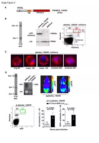

Analysis of PEXEL-Tagged Plasmodium berghei Schizonts Using mCherry and GFP Markers
E N D
Presentation Transcript
G1 G2 Hoechst GFP Supp Figure 5 A PEXEL PBANKA_136550 327aa mCherry TM B pbanka_136550::mCherry pbanka_136550 ::mCherry ANKAwt Chr 13- G1 G2 95%(1%) anti- mCherry ~100kDa Hoechst Chr 7- Uninfected rbc ~70kDa ctrl 3’utr dhfr mCherry C pbanka_136550 ::mCherry schizont 20h schizont 24h ring 4h troph. 16h troph. 12h D Chr 13- Δ pbanka_ 136550 ANKAwt 1x10⁷ 5x10⁶ ~1,5kb IBIS1 Chr 7- ctrl Chr 3- Δpbanka_136550 3’utr dhfr Δ pbanka_136550 K173cl1-GFP-Lucschiz -a Δ pbanka_136550 P<0.0001 P<0.0001 GFP+-schizonts (%) – G2 all schizonts (%) – G1 Hours post infection